Quantcast
Channel: VIETNAMESE MEDIC ULTRASOUND
Viewing all 624 articles
Browse latest View live

CASE 286: HCC GOING TO HEART, Dr PHAN THANH HẢI, MEDIC MEDICAL CENTER, HCMC, VIETNAM

$
0
0
MAN 46 yo, chronic hepatitis B, abdomen distension and dyspnea (photo).


Cardiac  ultrasound   showed  the  mass  intra  right  atrium  from  IVC (SEE  ECHO CARDIO 3D).


 ABDOMEN US FOUND OUT  LIVER TUMOR  WITH  BIG THROMBUS  INTRA IVC  EXTENSION TO RIGHT ATRIUM.


MSCT  WITH  CE   ALSO   DETECTED  THROMBUS  INTRA  CARDIAC  AND IVC.


BLOOD TESTS:   HBV POSITIVE  AND  AFP  1500 UI/ML.

CONCLUSION...HCC  FROM  THE  LIVER  GOES TO IVC  UP TO  RIGHT ATRIUM.

REFERENCE:
 .

CASE 287: CAROTOID PSEUDOANEURYSM. Dr PHAN THANH HẢI, MEDIC MEDICAL CENTER, HCMC, VIETNAM

$
0
0
Man  57 yo  detected  one mass at right  neck in pulsation.

Ultrasound  first at one clinic reported that  was a pseudoaneurysm  of  right  common carotid with thrombus inside  ( because  it had color inside this mass on  doppler).( see us 1, us 2).


Ultrasound  review at MEDIC:   it is a cystic mass  of  right lobe  thyroid gland without   Doppler  flow inside)  See  ( image us2, us4)


Reference from doctor  requested  CT angio of carotid  artery to make  sure  diagnosis of normal  carotid artery.


FNAC  was done for this thyroid mass which was to be a colloidal cyst.


What is atefact on color Doppler to make the misdiagnosis at the first ultrasound?
What is setting  parameter of  ultrasound color Doppler for elimination  this atefact?

CASE 288: BLACK SKIN TUMOR, Dr PHAN THANH HẢI, MEDIC MEDICAL CENTER, HCMC, VIETNAM

$
0
0
Woman 55 yo  worried about the  black skin spot at the left eyebrow  which was getting bigger and  itchy recenlty in this month ( see foto),  and one dermatologist made a caution that was to be a melanoma.


Ultrasound  of this skin tumor by 15MHz probe with Doppler showed that was a superficial skin tumor, size of 2 cm  and rich vessel supplying.
Elastoscan ultrasound  showed this tumor in blue code, not invasive to deep layer.


What is your diagnosis for it ?


Operation for resection this tumor and microscopic report was a pigmented papilloma with melanocyte (see  pathology report).

CASE 289: GRAPE'S SIGN in LIVER, Dr PHAN THANH HẢI, MEDIC MEDICAL CENTER, HCMC, VIETNAM

$
0
0

Man 27yo  one  month  ago  pain at  liver  with  low grade  fever.
First visiting of MEDIC ultrasound  liver  was detected  at  segment 7 of liver  multiple  spots,  hypoechoic size of 1-2 cm  with  the round border,  no  rising vessels on Doppler. Elastoscan this lesion was soft  tissue  as  an abscess. 


 Blood test presented  WBC rising and parasite tests were  normal.


One  week later  with  more pain  at liver,   CT scan with CE made image  like  grapefruit   as  an abcess.
Blood tests:  WBC rising  and  more eosinophil, CRP and IgE rising.


What is  your  diagnosis for the case?.

CASE 290: PARACERVICAL SPINE ABSCESS: Dr PHAN THANH HẢI, MEDIC MEDICAL CENTER, HCMC, VIETNAM

$
0
0
Woman 30 yo presented cervicalgia and swelling of the neck.


Chest Xrays: one mass at right of her neck.


Ultrasound detected thyroid gland normal( US1), but retrothyroidal spaces both 2 sides had hypoechoic masses, no vessels  intra mass which were suspected abscesses ( see  US 2:cross-section  at  lower  part of the neck, and US 3:longitudinal scanning of the neck).


MSCT of the neck (CT1 image showing the normal thyroid gland and 2 mass at lateral cervical spine looked like abscess). Frontal  view  and  sagital view detected  osteolytic lesions  at thoracic spine T1.


Ultrasound guided aspiration pus from abscess and detected BK positive in pus analysis.

It was a POTT' abscess of the neck, due to spine T1 tuberculosis.

CASE 291: ABDOMINAL PAIN POST CESAREAN, Dr PHAN THANH HẢI, MEDIC MEDICAL CENTER, HCMC, VIETNAM

$
0
0
Woman  34 yo, very painful some days during menstrual cycle for 2 years ago and after  cesarean operation  3 years before. Pain at  10cm  upper  pubis   near middle line and clinical palpation detected  one  mass of round shape and pain  at pressure.
Ultrasound  scan detected this mass ellypsoid, size 4cm x 3 cm intra right  rectus abdominus  muscle.
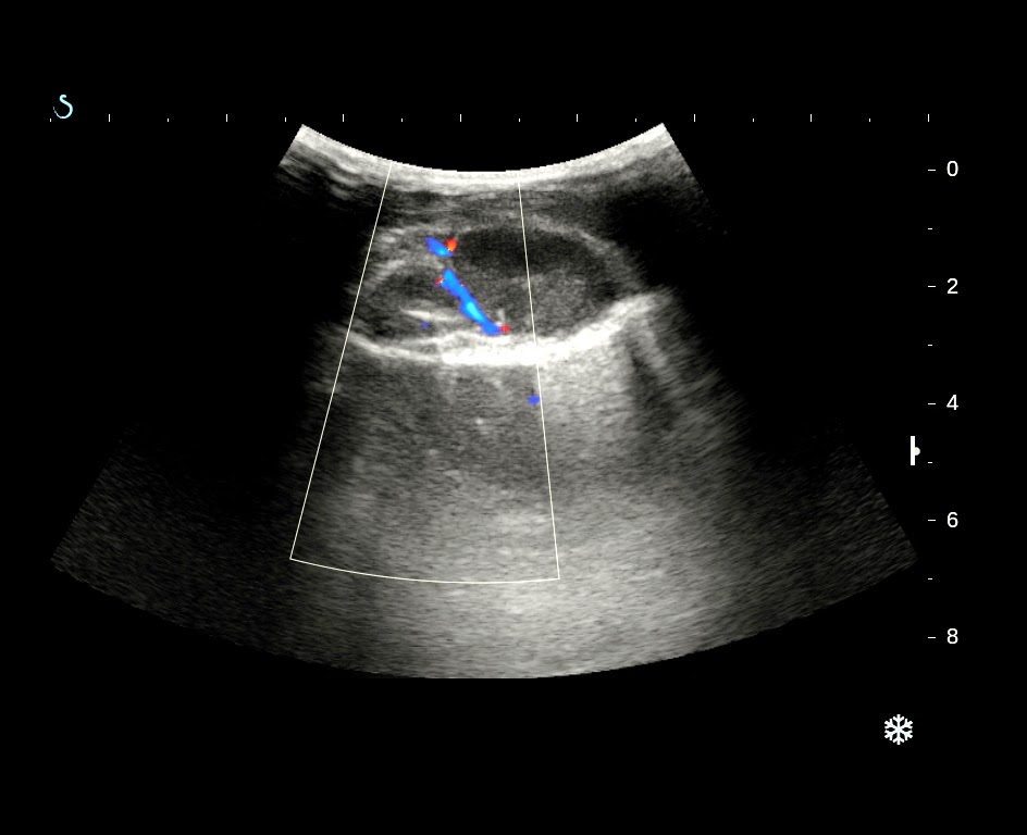
With  probe  3.5 MHz  this  mass was hypoechoic pattern looked like a cyst,  with  a small artery inside (see image 1, and video).


With linear probe 15MHz  this mass  had inhomogeneous structure, central  necrosis. Elastoscan this mass was  soft at central part (see  image 3 and 4).


FNAC was done for diagnosing the mass. Result from pathologist was hematoma with cells that were suspected endometrium cells.


What are your guidings for treatment for the patient?

CASE 292: BREAST TUMOR of a YOUNG GIRL, Dr PHAN THANH HẢI- Dr JASMINE THANH XUÂN, Dr HỒ CHÍ TRUNG, Dr TRẦN THỊ HỒNG VÂN, Dr VÕ ĐĂNG HÙNG, Dr NGUYỄN SÀO TRUNG, Dr HỨA THỊ NGỌC HÀ, MEDIC MEDICAL CENTER, HCMC, VIETNAM

$
0
0
15yo patient from Dong thap province. For one month there was a small lump which was about one finger at right breast but it was getting bigger rapidly, and painless.


Right breast mass presented hypoechoic pattern, nearly took entirely breast volume, # 8cm x 10cm, hypervascular. Upon breast US the mass was inhomogenous, increasing diastolic phase and decreasing RI=0.37.


Mammo and MRI= Phyllod tumor or huge fibrosarcoma (BIRADS 3-4).


Core biopsy and immunohistostaining result: Malignant phyllod tumor or sarcoma of myxoid cellular stroma.


Radical mastectomy was done. Microscopic result was poor differentiated sarcoma.


Reference


CASE 293: KIDNEY ABSCESS, Dr PHAN THANH HẢI, MEDIC MEDICAL CENTER, HCMC, VIETNAM

$
0
0
Woman 58yo diabetes for 10 years, fever and epigastric pain radiating to left flanc.
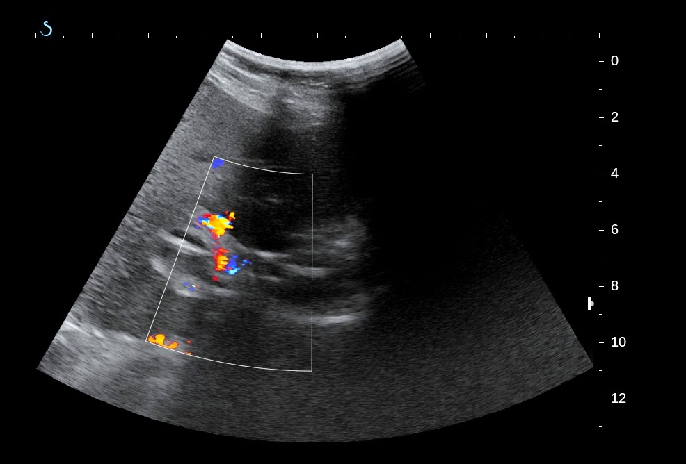
Ultrasound of abdomen first detected one mass with air inside nearby left kidney which made bright line artifacts (see video clip), so that was thought to be a renal abscess.


MSCT with CE detected the abscess with air inside destroying lower pole of left kidney.


Blood test with WBC rising very high= 23,760 /mm3 with neutrophil 81.5%  and glucose urine=56mmol/L.


Ultrasound guided punction of renal abscess removed white pus. 


Bacteriology analysis result was  gram negative bacilli.
Treatment:antibiotic per IV  and surgical drainage of renal abscess and controlling  blood sugar by insulin.
Summary: Kidney abscess of diabetic patient was detected by ultrasound in recognizing gas inside abscess.

CASE 294: SIGMOID COLON VOLVULUS, Dr PHAN THANH HẢI- Dr LÊ THỐNG NHẤT, MEDIC MEDICAL CENTER, HCMC, VIETNAM

$
0
0
Male patient 56 yo,  in emergency  going to MEDIC  by  acute  abdominal pain  and  distension. Clinical examination   this  patient cannot lay down ( see foto)
Ultrasound scanning of abdomen  first  detected  colon distension with air  and  hyperperistaltism (see 2 US images).


Next step  a  standing xrays of abdomen was done  with  the  sign C-loop, typical of  sigma colon  torsion (see   xrays plain film).
MSCT of abdomen without CE presented  dilated colon  with air   (CT 1 double  black  ring  of  colon sigma distention,   CT 2 image  section of sigma colon  asymmetric,  CT3:   image of coffee  beam, CT 4 : frontal section  with  mesocolon in torsion).
Radiologist  reported   volvulus of  colon sigma for the case.
Emergency  surgery detected  one part of  sigma colon  ischemic, resection  and colostomy with double canon technique.
Conclusion:  Emergency case  with ultrasound first  choice for diagnosis, conventional x-rays  also can help patient  but  CT is the  best  information for this case.

CASE 295: SPLEEN TUMOR, Dr PHAN THANH HẢI, MEDIC MEDICAL CENTER, HCMC, VIETNAM

$
0
0
Woman  63 yo, pain at  left subcostal  for one  month without  fever.

Abdomen ultrasound detected  the  spleen changing its surface, irregular lobular border with  many hypoechoic  structures  intraspleen  from hilus and free fluid  around the spleen ( see us 1, us 2).


MSCT with CE found out   inhomogeneous structure of spleen, with  many  hypodense zones, non enhancement  with contrast  from  hilus of  spleen  radiated toward peripheric zones of  spleen,  and  tail of  pancreas was adherent to spleen hilus.

Radiologist  suggested tumor  of  the tail  of pancreas invasive to hilus of spleen ( see  ct 1, 2, 3).


Blood tests were normal all cancer markers, and blood amylase highly elevated.


Preoperative  diagnosis  the case  was  vascular thrombosis of  spleen due to  inflammation of the pancreatic tail.
Operation  for  splenectomy, and  removing the hilus mass of spleen ( see macro).


Microscopic report  was chronic necrosis due to inflammation.

Discussion: Clinical  with pain for more one month  at left upper adominal  area whch was  KEHR' s sign.

Ultrasound detected  many avascular zones  in spleen.

MSCT with CE  find out  wedge – shape.
Blood test : high  amylase, looked like  PANCREATITIS  at the tail  complicated to hilus  of SLPEEN  INFARCTION.


REF  case report  from  JOP.


CASE 296: TUMOR of TESTES, Dr PHAN THANH HẢI-Dr NGUYỄN MINH THIỀN, MEDIC MEDICAL CENTER, HCMC, VIETNAM

$
0
0
Man 55 yo  pain at left  scrotum some  months ago and himself  detected  one mass in  left scrotum.

One  urologist  in palpation  it  suggested 2 testes in left  scrotum.
Ultrasound  scanning of  left  scrotum  showed that a small amount fluid   hydrocele vaginalis  and  one mass  rounded- border  in  adherence to  left testis with  size of 4 cm, This mass   was  hypovascular  and  soft upon  ultrasound elastoscan (see  3 ultrasound pictures of  this mass).


MRI  with gado of the mass presented it from epididymis that was enhanced with  contrast  gado and suggesting an ADENOMATOID  TUMOR of epididymis.


It is a benign tumor, biopsy was done  and macroscopic report was in correlation to imaging diagnosis.


Operation for removing the mass and microscopic confirmed the imaging diagnosis again.  

REFERENCE:


CASE 297: CHRONIC BOWEL OCCLUSION, Dr PHAN THANH HẢI, MEDIC MEDICAL CENTER, HCMC, VIETNAM

$
0
0
Man 47yo,   history of  3 months  with  peristaltism crisii and severe pain at  RLAQ more and more.
Ultrasound  first showed that  small bowel  dilating in  hypermotion and ascites.


MSCT  detected  enterolysis, stenosis at ilium  pre-cecum and suspected tuberculosis. 


Medical  antituberculosis treatment was in setting up, but  onset  total  occlusion was  acute  in emergency.
Operation in emergency detected  one portion of  ilium in hard stenosis. 


Resection that portion and  microscopic result  is adenocarcinoma infiltrating the bowel.

CASE 298: Carcinomatosis Ascites, Dr PHAN THANH HẢI, MEDIC MEDICAL CENTER, HCMC, VIETNAM.

$
0
0
Woman  64 yo, abdomen distention slowly for 3 months (photo).


Ultrasound  first  found out ascites  with slouge fluid  and  many debris, normal liver, omentum thickening  like cake with  many hypoechoic nodules  and pelvis no tumor.


Chest X-Rays  was  normal.


MSCT of abdomen reported  a large amount of  ascites and  great omentum  thickening  with  many  nodules,  enhanced with  CE  and no  ovary  tumor.


Punction of  yellowish  ascites  that cytology was  negative and  ADA negative. Blood test  was  very high CA 125.


Laparoendoscopy cannot detect  primary tumor, but many white  nodules  covered  the  great omentum  but not in parietal peritoneum.
Biopsy the great omentum nodule. Microscopic report  was  undiffentiated adenocarcinoma, suspected  come from GI TRACT or  OVARIAN CARCINOMA.


What is  your  interpretation?.


CASE 299: PORTAL VEIN FOREIGN BODY, Dr PHAN THANH HẢI, MEDIC, MEDICAL CENTER, HCMC, VIETNAM

$
0
0
Woman 65 yo, epigatric pain for one week, cannot eat  and  no fever.
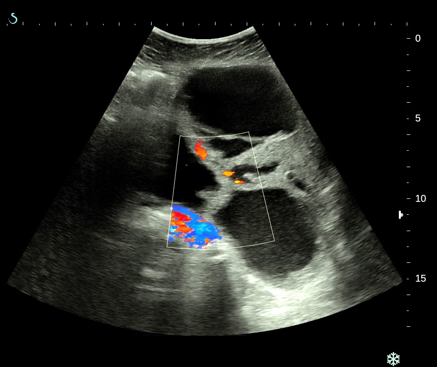
Ultrasound of abdomen  in decubitus position  detected  vena porta thrombosis and some  white lines intra portal vein which  came from the wall of gastric  antrum (see 4  ultrasound  pictures  in ventral view).


For clear viewing of  portal  vein  we  scanned  the liver  by sitting position and dorsal view.


Portal vein  was in distension, no flow  due to  thrombosis, and  in crossed section of portal vein we detected a white foreign body.( 2  pictures  with  sitting position scan ).

MSCT with CE  for  evaluation portal vein found out  the  foreign body which  length of 5 cm  intra left  branch of portal vein and one another end was intra gastric antrum wall.
The foreign body was  covered by thrombosis intra  left branch of portal vein (see 3 CT  images).


Blood tests  confirmed  infection  with  rising WBC and high CRP, no  abnormal coagulation test.


With  the  past history of ultrasound  scanning in ventral and dorsal views, MSCT and blood tests, the first choice  of diagnosis was intraportal vein foreign body, which was liked toothpick in penetration the gastric wall and  entering  liver  to left branch of portal vein, that caused  portal vein thrombosis.

What is your suggestion and planning of treatment for the female patient?

FEEDBACK=

An anouncement about case 299 of MEDIC  on Google web after the case was posted  for 30 minutes.


CASE 300: MULTIPLE BONE TUMORS, Dr PHAN THANH HẢI, Dr HỒ CHÍ TRUNG, Dr LÊ THÔNG LƯU, MEDIC MEDICAL CENTER, HCMC, VIETNAM

$
0
0
XIN DOWLOAD THEO LINK ĐỂ THẤY HÌNH

case-300-multiple-bone-tumor

MAN 46 YO PAIN AT LEFT LEG  AT PALPATION. X-RAYS  DETECTED TIBIAL BONE EROSION.


ULTRASOUND  SCAN THIS MASS SHOWED CYSTIC FORMATION.( SEE US  PICTURES)


CHEST XRAY  FOR ROUTINE CHECK UP  PRE OP  DETECTED  ONE MASS AT  RIGHT THORACIC WALL..( CHEST XRAY).


ULTRASOUND SCAN THIS MASS WAS  SOLID MASS,  HYPOVASCULAR WHICH WAS FROM THE  RIB. (SEE US3..WITH 3.5 MHz,,US 4 WITH 12 MHz, US 5 CDI,  US 6  ELASTOSCAN).


MSCT of  THORAX: THIS  MASS WAS  FROM  THE RIB   AND  ANOTHER  MASS AT  RIGHT  LUNG SUSPECTED  CANCER.


BLLOD TEST REPORT=   CYFRA -21  HIGH.


CORE  BIOPSY  OF THORAX WALL MASS = METASTASIS TUMOR HISTO TYPE  ADENOCARCINOMA    SUSPECTED  FROM THE LUNG CANCER.


CASE 301: SKIN METASTASIS, Dr PHAN THANH HẢI, MEDIC MEDICAL CENTER, HCMC, VIETNAM

$
0
0
XIN DOWNLOAD THEO LINK ĐỂ THẤY HÌNH
FOR PICTURES  PLS DOWNLOAD LINK BELOW.

MAN 83 YO HAD BEEN  TREATED  LUNG CANCER WITH  CHEMOTHERAPY  AND RADIATION  FOR 2 YEARS.
ONE  MONTH  AGO  HE DETECTED  SMALL SKIN TUMOR  AT LEFT TEMPORAL SCALP  RAPID  GROWING AND BLEEDING ( SEE PICTURE).


ULTRASOUND  SCAN   REPORTED  THIS TUMOR BEING  FROM THE  SCALP NO  INVADE  TO  BONE ( SEE  2 US  PICTURES)  AND   LIVER  METASTASIS ( US PICTURE 3)


MSCT  CONFIRMED THE  SCALP TUMOR   NOT ERODING THE BONE, MANY  METASTATIC  LESIONS FROM THE LUNG TUMOR  TO  BOTH SIDE OF LUNG, LIVER, ADRENAL GLAND.


BIOPSY OF THIS TUMOR  CONFIRMED THAT  METASTASIS FROM LUNG CANCER,  SQUAMOUS CELL CARCINOMA..


SUMMARY:   LUNG CANCER  SOMETIMES  METASTASES TO SKIN OF SCALP AND NOT TO BONE.

REFERENCE:


CASE 302 : OVARY TUMOR , Dr PHAN THANH HẢI, MEDIC MEDICAL CENTER, HCMC, VIETNAM.

$
0
0
FOR PICTURES PLS DOWNLOAD THE LINK


Woman 50 yo, vaginal bleeding.
Pelvis ultrasound  detected  one mass  cystic, multiloculated   with  septation thickening and  solid part. No ascites (see  5 US pictures).


MRI  with  gado reported  with  enhanced  CE  suspected  ovary cancer.


Blood test :  CA125  rising  125 U/mL
Pre operative  diagnosis  is  ovary cancer  stage IIB. Microscopic  specimen report is  serous cystadenocarcinoma.


Discussion: With 3 modalities for diagnosing  this case ULTRASOUND, MRI  and BLOOD TEST MARKER, what  is  the value?.

CASE 303: INGUINAL SCARPA TUMOR, Dr PHAN THANH HẢI, MEDIC MEDICAL CENTER, HCMC, VIETNAM

$
0
0
FOR PICTURES PLS DOWNLOAD THE LINK


Woman  61 yo  detected  at right inguinal  scarpa a  small tumor  slowly  growth, no  painful,  size of  about  one egg. Clinical palpation was  subcutaneous, hard nodule,  fixed  to  deep  layer.
Ultrasound scanned it was  a round mass , size of 5 cm in diameter,  well-bordered, inside  structure  was  solid and inhomogeneous  ( US picture 1).


 Doppler  scanning  detected  hypervascular  with  hilus,  vascular supplying  of a lymph node (US picture 2), and   Doppler spectrum  of  hilus artery  was  pulsatile , V1= 52 cm/s and  RI=0.6.


Elastography ultrasound  was a soft mass, inhomogeneous structure.


Sonologist  cannot  find another abnormal  lymph node at  another  side, and report  suspected  inflammation  lymph node.


Blood tests  WBC, CRP, beta2 microglubuline  were  normal.


Core biopsy with immunohisto staining report was lymphoma.


Reference ;


CASE 304: HIP PAIN, Dr PHAN THANH HẢI, MEDIC MEDICAL CENTER, HCMC, VIETNAM.

$
0
0
PLS DOWNLOAD THE LINK FOR PCTURES


Man 43 yo, one week ago, pain at  his left  hip  while moving his thigh, no trauma history.
Ultrasound first saw  the  left hip joint  having a small amout of fluid collection  and  more  Power Doppler in muscles  around the hip ( see 4 US images).


MSCT was done but radiologist said nothing abnormal detected.


MRI detected abnormal head of left  femur  bone  looked like  aseptic necrotic femoral head.


Please compare  diagnostic values of 3 diagnostic modalities for this case:ultrasound, MSCT and MRI.

CASE 305: Presacral Tumor, Dr PHAN THANH HẢI, MEDIC MEDICAL CENTER, HCMC, VIETNAM.

$
0
0
FOR PICTURES PLS DOWNLOAD THE LINK


Man 65 yo in trouble  and difficult mictation, looked like in prostate disease.
Ultrasound of pelvis  detected  one presacral ovoid mass causes  deplacement  of rectum and  prostate (US picture 1).


Pelvis MSCT of this tumor presented solid, retrorectum, and presacral, well bordered, and not connected to rectum or  sacrum (see CT1, CT2).


MRI with gado of this  tumor is  central necrosis , well  enhanced  with gado contrast ( see  MRI 1, 2 ,3).


Blood tests detected abnormal anything.
What is your  suggestion for diagnosis?.


Viewing all 624 articles
Browse latest View live