Quantcast
Channel: VIETNAMESE MEDIC ULTRASOUND
Viewing all 624 articles
Browse latest View live

CASE 306 : THYROID or PARATHYROID TUMOR,Dr PHAN THANH HẢI, MEDIC MEDICAL CENTER, HCMC, VIETNAM

$
0
0
PLS DOWNLOAD THE LINK for PICTURES


 Lady 24 yo, 5 years  before  fracture of  left femoral head, and  now fractures of  2 bones of  right forearm by  falling trauma [see  photo].


X-rays  of  pelvis bone  made  pointed  osteoporosis of bone .


For screening, ultrasound of the neck  detected one ovoid mass, size  of  3-2cm,  hypoechoic  at the  lower pole of  the thyroid gland,  and  hypervascular on Doppler.


Sonologist suggested PTA for the case.
Osteogram BMD showed very lower bone  index.


Blood tests  =   PTH  very high and elevated  calcium.


Do you  make first choice of diagnosis of  PTA?

OPERATION  of  RIGHT  LOBECTOMY.THIS TUMOR WAS  WELL BORDERED, SOFT TISSUE.  ( see MACRO1,2).


MICROSCOPIC REPORT  WAS  PARATHYROID ADENOMA.


REFERENCE  


CASE 307: EXTRAPERITONEAL RUPTURE of URINARY BLADDER, Dr LÊ VĂN TÀI

$
0
0
FOR PICTURES, PLS DOWNLOAD THE LINK


A 38-year-old man has fallen motorcycle one month ago. About two weeks he had dysuria and bloody urination.

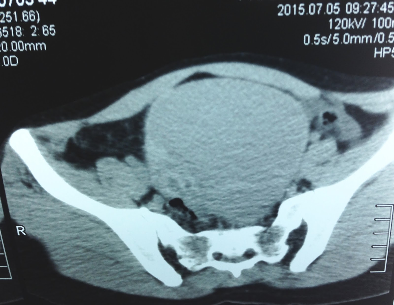
Abdominal ultrasound scans detect structure echo free with thickening border due to inflamed fat close to urinary bladder. After to rule out appendicitis, abscess and fluid collection of acute pancreatitis, color Doppler with grade-compressible technique was used and to reveal color flow of jet or stream from fluid-filled structure into urinary bladder. Therefore diagnosis of extra-peritoneal urinary bladder rupture was established.

Late phase contrast enhanced-MSCT showing urine sac close to urinary bladder.
There is urine accumulation adjacent urinary bladder at cystography. 
Patient has been operated promptly.

Key diagnosis of fluid sac due to extraperitoneal urinary bladder rupture:color Doppler flow of urine jet through gap from fluid sac into urinary bladder.   


CASE 308: PATELLA DISLOCATION, Dr PHAN THANH HẢI, MEDIC MEDICAL CENTER, HCMC, VIETNAM

$
0
0
FOR PICTURES PLS DOWNLOAD THE LINK


WOMAN 53 YO, CHIEF COMPLAINT  OF   SWELLING  AT HER LEFT KNEE, INSTABILITY AT WALKING. DIFFICULT SITTING AND STAN DING DUE TO  ONE MASS  MOVING  FROM  MEDIAN SITE  TO LATERAL SITE OF THE LEFT KNEE ( SEE FOTO THE LEFT KNEE SITTING  AND STANDING).


ULTRASOUND SCANNING  OF THIS  MASS  WAS FLUID COLLECTION  AND CANNOT DETECT  PATELLA BONE AT THE MIDDLE SITE OF THE JOINT (SEE 3 US PICTURES).


X-RAYS OF THE  LEFT KNEE  JOINT  MADE  DIAGNOSING  OF PATELLA  BONE DISLOCATION, MOVING  FROM  MEDIAN TO LATERAL  OF  MIDDLE LINE OF  THE KNEE JOINT ( SEE  3  X-RAY FILMS).


REFERENCES:


CASE 309: RENAL TRANSPLANT, Dr PHAN THANH HẢI, MEDIC MEICAL CENTER, HCMC, VIETNAM

$
0
0
FOR PICTURES PLS DOWNLOAD THE LINK


Male patient 76 yo,  with right  renal transplant 7 years ago, now he detects urine volume lesser than  500ml for 24 hrs, and nephrologist  requested  ultrasound screenning of  greff  kidney  in urgency and blood tests for kidney investigation.

Ultrasound found out  the  kidney greff at  right pelvis, size of 13cmx 9 cmx 7.5cm,   echo structure  liked normal, no  hydronephrosis,  and color Doppler   study of  the  vascular supply for this kidney  is  good   with  RI = 0.71.


What is your  suggestion  after  ultrasound examination for the case?.
 BLOOD TEST RESULTS=  NORMAL RENAL FUNCTION.

REFERENCES: 
ref 1 , ref 2


CASE 310: CAMEL HUMP'S SIGN of THE NECK, Dr PHAN THANH HẢI, MEDIC MEDICAL CENTER, HCMC, VIETNAM

$
0
0
FOR PICTURES PLS CONNECT 3G or  DOWNLOAD THE LINK


case-310-camel-humps-sign-dr-phan-thanh-hai


Woman 31 yo,   4 months ago detected  left postero-lateral of the neck swelling, no painful  (see foto).


XRays of the neck:  normal cervical spine bone (xray1, xray 2).


Ultrasound  of this mass  revealed  intra trapezius muscle  echo  very poor, hypovascular  crossing  middle line of the neck ( us 1, us 2).


MRI of  the neck  with gado, found out this mass  intra trapezius muscle  well- bordered and  enhanced with gado, and central necrosis.


What is your suggestion for diagnosing the case?.

Core biopsy was done. Wait for histoimmunostaining report.

CASE 311 : ACUTE HEMIFACIAL PAIN, Dr PHAN THANH HẢI, MEDIC MEDICAL CENTER, HCMC.

$
0
0
FOR PICTURES PLS CONNECT 3G or DOWNLOAD THE LINK 

Boy 14 yo, 7 days ago, pain at left  hemifacial and  fever.


ENT doctor  after  rhinoendoscopy said, but  one week with treatment  no response.

Emergency consultation  with  ultrasound  paranasal sinus ( us 1 imge= transversection scan  with 3.5 MHz laydown position at left  maxillary, showing  the sinus filling  with fluid /  us 2 imge=  at  right  sinus no fluid  only intrasinus air/  us 3 image=   in  comparison to  image  of sinus  scan ultrasound of R and L  maxillary / us 4 image = scanning in  sitting position ).


Ultrasonologist  suggested  acute  sinusitis of  maxillary.

MSCT  confirmed  this US diagnosis.


Blood tests  also  make  sure an acute infection.


Summary of case 311: quickly  scaningn ultrasound  at  paranasal sinus  make  exactly diagnosis  acute  sinusitis  in  emergency room.

REFERENCE:

CASE 312: FETUS in CONTRACTION IN UTERO: Dr JASMINE THANH XUÂN, Dr TÔ MAI XUÂN HỒNG

$
0
0
A parturient in 27 weeks of gestation, PARA 2002, could have not feelings  of her baby movement and went to OB examination for help.
Sonologist realized fetus in crisii of contracture with normal heartbeat and MRI was done to confirm this abnormal phenomenon of fetus.
Amniotic fluid analysis and karyotype and 22th chromosome were done.


What do you think about this case and what would you do in next step ?

CASE 313: FISH BONE APPEARING in NECK, Dr PHAN THANH SƠN, Dr NGUYỄN ĐỨC DUY LINH, Dr NGUYỄN NGỌC XUÂN GIANG, BÌNH AN HOSPITAL, KIÊN GIANG

$
0
0
FOR PICTURES PLS CONNECT TO 3G / DOWNLOAD THE LINK

case-313-fish-bone-appearing-in-NECK

Male patient 79 yo had foreign body on right neck (see pictures).
HISTORY
4 days before of hospitalization, during dinner with stingrays, in sneezing, a 79 yo patient took out and in some fish parts, and then feeling caught something in his throat but still eating.
Morning after he went to hospital as felt felt like a head of bone at his right neck.

ULTRASOUND

 Ultrasound at right neck revealed a fish bone in soft tissue, echogenic line in subcutaneous edema around the fish bone, but no vascular.

ENT ENDOSCOPY


 ENT endoscopy detected edema of right piriformis recess due to fish bone perforating outside from the throat.

CT SCAN
CT–Scan confirmed the fish bone in the right neck, not injured the right vessels of the neck.


The fish bone was removed out of the right neck, with 2.8 cm long of size.


Fish food is very interesting, but sometimes lead to dangerous situation like the case, specially for old patient. 

REFERENCE;


CASE 314:CALCIFIED LIPOMA of CALCANEUS TENDON and SOLEUS MUSCLE - - Dr PHAN THANH HẢI- Dr LÊ THANH LIÊM- Dr Nguyễn Đức Duy Linh; Dr Nguyễn Ngọc Xuân Giang – BÌNH AN GENERAL HOSPITAL_KIÊN GIANG_VIỆT NAM.

$
0
0
FOR PICTURES PLS CONNECT TO 3 G / DOWNLOAD THE LINK

case-314-lipoma-calcification-of-calcaneus-tendon-and-soleus-muscle-

A 30 year-old woman with history of mass within the right heel region 2 year ago, no pain  and normal walking.
ULTRASOUND
Ultrasound found anechogenic mass in the calcaneustendon and soleus muscle, hypovascular.
The surface of the mass was close to the Achilles tendon, well-defined with the capsule. Deep surface was very difficult to survey. Intra the mass there was an hyperechoic structure with strong shadowing, like bone. Ultrasound did not determine that there was continuity between the mass with calcaneus tendon or not.
Sonologist suggested a diagnosis of chondro-osteoma and organized synovial cysts of the ankle. Dx: Lipoma calcification.

CT SCAN
MSCT detected a lesion in the soleus muscle, density of fatty tissue, 10x4cm in size, around of achilles tendon, clear capsule, rounded border, with a very high-density nodule inside, looked like the bone. The lesion was not associated  with the calcaneus bone and the leg bone. Radiologist suggested a diagnosis of calcified lipoma.


OPERATION
Surgery was conducted in next day. Macroscopic image looks like the lipoma with calcified nodule inside.


Waiting for  microscopic results.

REFERENCE:

CASE 315: SPONTANEOUS PORTO-SYSTEMIC SHUNT, Dr PHAN THANH HAI, Dr NGUYEN THI ANH HONG, Dr LE THONG NHAT, Dr TRAN LAM

$
0
0
FOR PICTURES PLS CONNECT TO 3G / DOWNLOAD THE LINK

case 315

Woman  63 yo,  5 years ago  had been treated  diabetes  not  control blood  sugar. Patient  has  some  subcoma  crisii and had been treated in many  hospitals, now she  is in somnolence, easy in sleeping  after eating but cannot sleep at night.
MRI of  the brain, radiologist  detected  hyperintense  T1 at basal ganglion area, susgested  hepato-encepalopathy (image MRI).


Checking  the liver by blood tests, liver function is still good,  but ultrasound of  liver  detected  porta-hepatic vein shunting  very high flow ( see US image 1 dilatation cystic  intrahepatic, US 2, color Doppler: Shunting porta-hepatic and US video).


MSCT angio of  liver with  3 phases: arterial phase shows that not abnormal venous  phase : dilated  the  porta-hepatic  anastomosis like snake. In 3D CT reconstruction  confirmed a  high flow porta-hepatic shunting).


Bood test reports  this time  no ceton , high NH3 = 88.89 micromol/mL (normal  18-72).
Conclusion: It is  a high flow  spontaneous  porta-systemic  shunting appeared as hepatic encephalopathy in clinical examination.

REFERENCE:

CASE 316:RETROPERITONEUM CALCIFACATED TUMOR, Dr PHAN THANH HẢI, MEDIC MEDICAL CENTER, HCMC, VIETNAM

$
0
0
 FOR PICTURES PLS CONNECT TO 3 G / DOWNLOAD THE LINK


WOMAN 45 YO..PAIN  AT  RIGHT  SUBCOSTAL REGION. PLAIN XRAY FILM  KUB DETECTED  CALCIFICATION  PUNCTIFORM   AT LIVER BORDER AND  RIGHT KIDNEY ( SEE XRAY PLAIN FILM).


ULTRASOUND  OF ABDOMEN  FIND OUT  THIS MASS  IN RETROPERITONEUM,  SIZE  OF 7X5 CM , WHICH PULL THE RIGHT KIDNEY DOWN. ITS STRUCTURE  IS  CYSTIC,  MULTILOCULATED WITH  FINE WALL  AND  CALCIFICATED. THE CONTENT OF THIS CYST IS  MIXED ECHOGENIC STRUCTURE ( SEE  4 US IMAGES ).


MSCT OF ABDOMEN  WITH CE=  THIS TUMOR IS  MULTICYSTIC  WITH  CALCIFICATION OF  THE WALL ( SEE 3  CT IMAGES).


BLOOD TESTS  AND  MARKER S = AFP,  BETA HCG ARE NORMAL

WHAT IS YOUR  DIAGNOSTIC SUGGESTION   AND WHAT YOUR TREATMENT FOR HER?

CASE 317:CT FIRST US SECOND IN CAUTION OF THYROID FUNCTION, Dr PHAN THANH HảI, MEDIC MEDICAL CENTER, HCMC, VIETNAM

$
0
0
FOR PICTURES PLS CONNECT TO 3G/ DOWNLOAD THE LINK


MAN 29YO  TRAUMA  AT THE HEAD AND NECK BY TRAFFIC ACCIDENT, NOW IN  PAIN  AND TACHYCARDIA.

CT  NON -CE WAS FIRST LINE, RADIOLOGIST  REPORTED    BIG AND  DARK THYROID GLAND , CT UNIT ARROUND   85 UI HOUSFIELD ( SEE  FOTO AND  CT1,CT2 ARE  ABNORMAL).


 ULTRASOUND  OF  THYROID  ALSO    REPORTED LARGE VOLUME WITH  HYPOECHOIC AND   VERY  HIGH  SIGNAL DOPPLER . SPECTRAL DOPPLER  OF  SUPERIOR THYROID ARTERY  WAS VERY  FAST PULSATILE, TYPICAL HYPERTHYROIDISM  AS  GRAVE  DISEASE (BASEDOW).


BLOOD TESTS  CONFIRMED  A  HYPERTHYROIDISM  WITH TSH  LOW  VALUE  AND VERY  HIGH T3 AND T4.


SUMMARY: CT  NON CE  IS  POTENTIAL IN CAUTION  THYROID FUNCTION.


CASE 318 : DIFFUSE LYMPHADENOPATHY, Dr PHAN THANH HẢI, MEDIC MEDICAL CENTER, HCMC, VIETNAM.

$
0
0
FOR PICTURES PLS CONNECT TO 3 G/ DOWNLOAD THE LINK

case-318diffuse-lymphadenopathy

Woman  56 yo  slowgraded  fever for  one month, swelling of the neck and left  supra clavicular area.
Ultrasound  the neckpresented   normal thyroid and  many lymph nodes  hypoechoic without  hilus nodes (US image )


Abdomen ultrasound scan detected  aorta  elevated  by  hypoechoic mass, para-retro aortic  just  prolonged to  pelvis (US 1, US 2) and sonologist  suggested  lymphoma.


MSCT  with CE  for  staging  the  mass  nodes   supreclavicular  left , mediastinal, para aortic   and  inguinal area..( CT 1, CT2, CT3) =  stage IV  LYMPHOMA


BLOOD TESTS  with  high  beta microglobuline and  ferritin.


Biopsy supraclavicular node   and  histo immuno chemistry  report were  LYMPHOMA  diffuse  large B CELL.


CASE 319: LINGUAL THYROID, Dr PHAN THANH HẢI, MEDIC MEDICAL CENTER, HCMC, VIETNAM

$
0
0
FOR PICTURES PLS CONNECT TO 3 G / DOWNLOAD THE LINK


WOMAN  34 YO  HAD BEEN SUFFERED FROM RECURRENT SORE THROAT, COMING TO SEE  ULTRASOUND OF THE NECK MANY TIMES.

SONOLOGIST  REPORTED  THAT CANNOT FIND  OUT THYROID GLAND  AT THE NECK. ULTRASOUND   CANNOT  FIND  THYROID GLAND NOR THYROID ARTERY (SEE  US1, US2).


BLOOD TEST  =  TSH  6,19UI (N 0,3-5UI)  FREE T4 :1,3 N (0,7-1,8).
MSCT  OF THE  NECK  NON CE   DETECTED  2 HYPERDENSE MASSES  SIZE OF 2-3 CM   WITH HU  119 AT THE BASE OF LINGUAL  AND  BUCCAL PLANTAR ARE (CT1 ).


MSCT  CE OF  THIS MASS =  VERY  QUICK  ENHANCE  TO HU 176 ( CT2).


CT3: LATERAL  VIEW  3 MASSES, CT4   FRONTAL VIEW , CT 5   VASCULAR  OF CAROTID  NO DETECTED  THYROID ARTERY, CT6  3D VIEW OF 2 MASSES.


CONCLUSION :ECTOPIC THYROID GLAND  AT  LINGUAL  AREA.


CASE 320: VENA PORTA THROMBOSIS BY HCC, Dr PHAN THANH HẢI, MEDIC MEDICAL CENTER, HCMC, VIETNAM.

$
0
0
FOR PICTURES PLS CONNECT TO 3 G / DOWNLOAD THE LINK


Woman 58yo, history of  infected  HBV  and diabetes. One week ago  she was very  painful  in  liver region.
Chest x-ray showed elevated  right diaphragm ( see Xray chest film).


Ultrasound  reported that  no pleural effusion..but  liver  had  many tumors with  portal vein thrombosis  completly..( us1, us 2, us 3, us4 and video).


Blood tests = HBsAg  positive  with  AFP = 9.3 ng/mL.


Blood tests  again  with   HCC risk  on Wako machine resulted  very high  level of DCP, this test made thinking  portal vein  thrombosis  by HCC.


Discussion : History of  infected  chronic HBV,  and normal..AFP,  but  ultrasound suggested   VP THROMBOSIS due to HCC  that based on  WAKO  compiling  3 tests  AFP, AFP-L3%, DCP which confirmed  HCC.

CASE 321: MALE BREAST TUMOR, Dr PHAN THANH HẢI, MEDIC MEDICAL CENTER, HCMC, VIETNAM

$
0
0
FOE PICTURES PLS CONNECT TO 3G/ DOWNLOAD THE LINK
/case-321-male-breast-tumor

Male 58 yo  history of sefl  detected  right  breast tumor  slowly growing for 10 years [see FOTO],  its size of  6 cm and  changing color of covered skin.


On ultrasound   it was a solid tumor  with cystic formation, hypovascular, no  adherence to deep  muscle layer and small  axillary nodes ( US 1, US 2, US 3).


Mammography  showed well- bordered  round tumor,   no calcification ( mammo1, 2).


MSCT  with CE of this  tumor was  low CE enhanced,  very clear border (CT 1,2,3).


FNAC report  was  blood cell only and no tumor cell.


Operation  removed this tumor ( 2 specemen )


Pathology report  was  caverneous  hemangioma.


CASE 322: OVARY TWISTING, Dr PHAN THANH HẢI, MEDIC MEDICAL CENTER, HCMC, VIETNAM

$
0
0
FOR PICTURES PLS CONNECT TO 3G / DOWNLOAD THE LINK
case-322-ovary-twisting-

WOMAN 36 YO, 3 DAYS  AGO PAIN AT  SUPRAPUBIS, AND POLYKYURIA.
ULTRASOUND  OF  PELVIS WAS NORMAL.

PAIN CONTINUING  AND  RISING  AND SHE  WAS IN ADMISSION  OF ONE  EMERGENCY HOSPITAL,   IN CLINICAL SUSPECTED  RENAL COLICKY PAIN.
ULTRASOUND  AGAIN   DETECTED  ONE SUPRAPUBIS MASS,  SIZE OF10CM,  CYSTIC WITH  CLOUDY  FLUID INSIDE (US 1).


CDI  REPORT  WAS SMALL UTERUS, CANNOT  DETECT VASCULAR  AT  RIGHT UTERUS CORNER (US 2).


THIS CYSTIC MASS  WITH  MASS INTRA CYST  LIKED  SEBUM THAT  SUSGESTED  RIGHT OVARY TERATOMA  IN TORSION.
NO STONE  INTRA URINARY SYSTEM.

MSCT  NON CE   PRESENTED  OVARY MASS  LOOK LIKED TORSION OF TERATOMA.


EMERGENT OPERATION BY LAPAROTOMY DETECTED THIS MASS BEING AN OVARIAN CYST   BLACK IN  COLOR DUE TO TORSION ( SEE FOTO)


OPEN  SURGERY  REMOVED  RIGHT  OVARY MASS  ISCHEMIA AND BLEEDING INSIDE OF A  TERATOMA TUMOR.


Discussion : Why the first ultrasound examination was  normal?  Looking  the first picture ultrasound of the uterus that was  clear view and  the fluid  over  look liked urinary bladder.
But  you can see the small urinary bladder  in the lower corner of the first ultrasound picture.
The mistake  was due to ultrasound  scanning  with  the urinary bladder  not full filling , the ovary cyst  was not  septation because scanning view in small window.

The ultrasound examination is better  view  with  lateral decubitus position, but in some cases cannot see well in decubitus position.

CASE 323: ELEPHANTIASIS, Dr PHAN THANH HẢI, MEDIC MEDICAL CENTER, HCMC, VIETNAM.

$
0
0
FOR PICTURES PLS CONNECT TO 3 G / DOWNLOAD THE LINK
/case-323-


BOY 16 YO, HISTORY OF    ABNORMAL DETECTION OF HAND AND LEG IN THE CHILDHOOD, SLOW GROWTH ( FOTO).


2 HANDS  AND RIGHT LEG ARE ABNORMAL. PALPATION OF THE SKIN FEELS  EDEMA AND COMPRESSIBLE, NO PAINFUL.
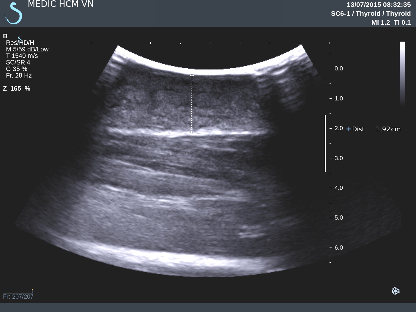
ULTRASOUND  of  RIGHT LEG  REVEALED  SKIN AND SUBCUTANEOUS  LAYER THICKENING  TO 1,9CM, NO RAISED  VASCULAR STRUCTURE.


MSCT  NON CE SHOWS  SKIN AND  SUBCUTANOUS   FATTY EDEMA, LOCATED THE  2 HANDS AND  FOREARM, RIGHT  HALF BODY, AND RIGHT LEG WHILE   BONES AND MUSCLES ARE NORMAL.


WHAT IS  YOUR  DIAGNOSIS?.

CASE 324: BREAST PSEUDOTUMOR, Dr PHAN THANH HẢI, Dr JASMINE THANH XUÂN, MEDIC MEDICAL CENTER, HCMC, VIETNAM

$
0
0

FOR PICTURES PLS CONNECT TO 3 G/ DOWNLOAD THE LINK


Woman 52 yo, history of hypertrophy of bilateral mammar gland. She had been in operation  for reduction of breast  size   10 years before. After operation she  detected  the  mass at the  site operation,  6- hour  position  at  2 breasts more and  more hard and no painful.
Ultrasound  check-up detected  at 2 breast,  masses  size  arround 5cm under  the skin incision at 6-hr position  of the breast,  hypoechoic  with  lateral shadowing.  At the left  breat mass  had  calcification,  soft elasto Q-score (see 5 ultrasound scan).


Mammography  cannot detected this mass.


MRI of breast without  gado, T1 ,T2 and fat subtraction, showed  fat  tissue with fibrosis  around and no axillary  lymph node.


Core biopsy of  this mass  and  reported  that  fatty tissue with  fibrosis, the border
was  fibrosis of scar  after operation  looked like a tumor. 


CASE 325: STRETCH MARKS SKIN, Dr PHAN THANH HẢI, MEDIC MEDICAL CENTER, HCMC, VIETNAM

$
0
0
FOR PICTURES PLS CONNECT TO 3 G / DOWNLOAD THE LINK


Boy 15 yo, history of  treated  high dosage  corticoid  due to uveitis;  2 years later  detected  many  scars  over  skin  at abdomen, leg, arm, back ( foto).

Ultrasound  of skin abdomen  shows  that  skin thin  and  hyperechoic, soft elasoscan ( see  US pictures 1,2,3,4).


Conclusion: It is  stretch marks skin due to  overuse  corticoid  which  destructed elastic fibers  in  derma layer of the skin.

REFERENCE:

Cases  of  skin stretch marks.


Viewing all 624 articles
Browse latest View live