A 31-year-old male patient complained about 3-day mild fever, right subcostal abdominal pain. He did not have any other symtomps such as voimiting, diarrhea and no history of abdominal surgery, trauma, liver biopsy or alcohol abuse. On physical examination, no mass in the right subcostal. B-mode ultrasound (US) findings showed a cystic structure (21x21mm) in the sixth segment, it communicated with 2 parallel –dilated - tubular - structure (d = 8 and 9mm) originated from the right portal vein and right hepatic vein. Doppler US showed yin-yang sign, right portal vein flow and right portal vein flow in the cystic structure. MSCT Angio comfirmed the AVM in right lobe of liver. The patient underwent an abdominal laparoscopic surgery for resection the AVM. The patient remains well postop. Wait for microscopic result.
↧
CASE 247: INTRAHEPATIC AVM, Dr PHAN THANH HAI, Dr NGUYEN CAO CUONG, Dr TRAN NGAN CHAU, MEDIC MEDICAL CENTER, HCMC, VIETNAM
↧
CASE 248: GASTRIC WALL TUMOR, Dr PHAN THANH HẢI, MEDIC MEDICAL CENTER, HCMC, VIETNAM
Woman 75 yo vomiting, endoscopy detected tumor extragastric fundus (see pictures).
Ultrasound of abdomen showed one hypoechoic mass with size of 4 cm, well-bordered at the hilus of spleen ( see 2 ultrasound pictures).
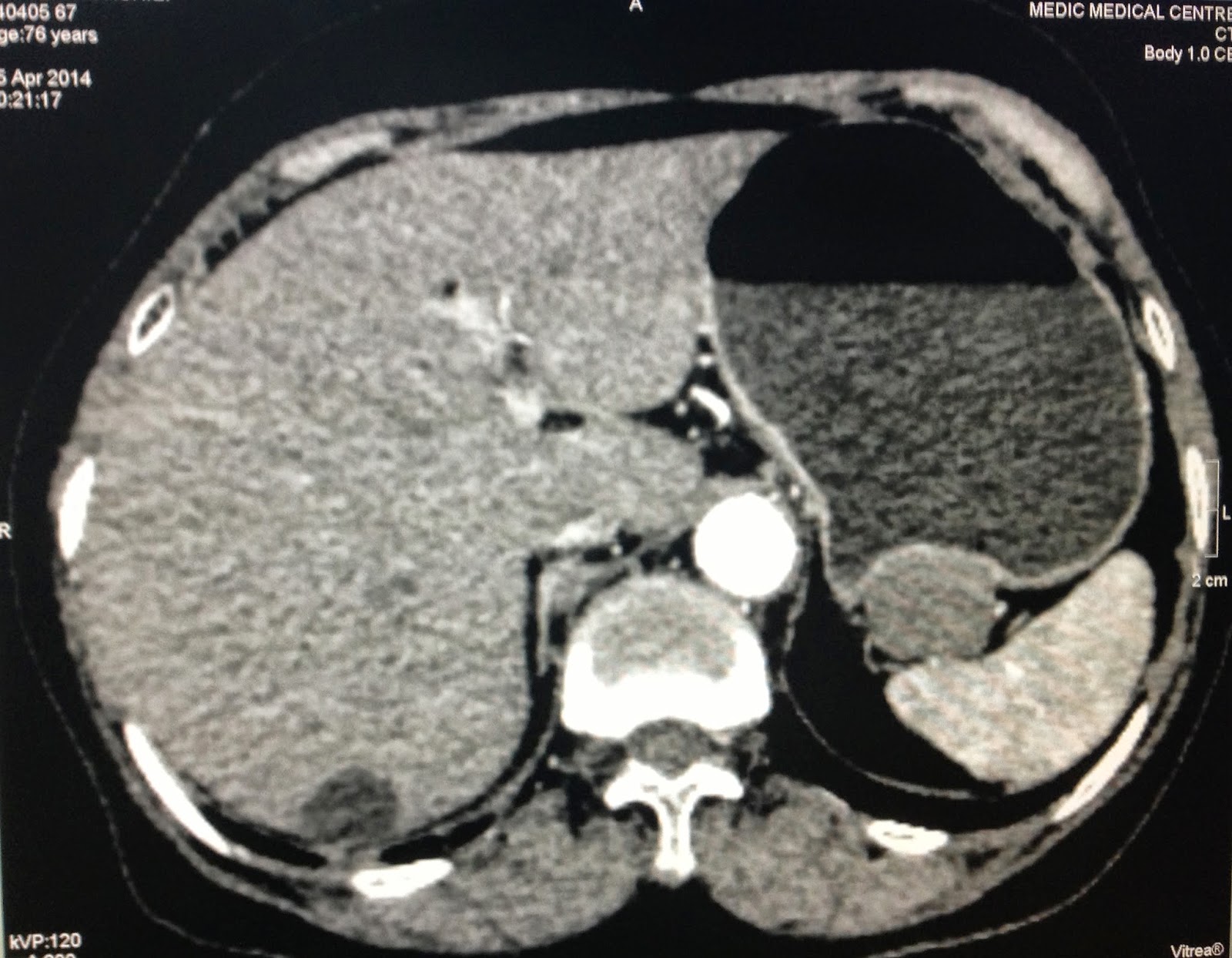
MSCT with CE found out this mass bending the wall of great curvature of stomach, very slow CE enhancement (see 3 CT pictures).
Blood tests of all markers are normal.
What is your suggestion of diagnosis?
↧
↧
CASE 249: RLAQ MASS, Dr PHAN THANH HẢI, MEDIC MEDICAL CENTER, HCMC, VIETNAM
Woman 56 yo, pain in RLAQ for 2 weeks, fever and GI tract trouble with diarrhea was treated by antibiotics.
Ultrasound detected one mass at RLAQ, suspected appendicular abscess or coecum tumor (see 4 US pictures).
Blood test WBC= 17K with 17% neutro.
MSCT with CE shows this mass being wall-off by instestine, central part is liquide as pus collection (see 3 CT pictures).
What is your suggestion of diagnosis and what is the another test needed for make sure diagnosis?.
↧
CASE 250: LOST of DENTAL PROSTHESIS, Dr PHAN THANH HẢI, MEDIC MEDICAL CENTER, HCMC, VIETNAM
Man 68 yo (photo) after drinking a lot of alcohol, he detected lost of dental prosthesis and dysphasia and apnea, like cardiac ischemia.
He came seeing cardiologist for dissolve his cardiac problem, but it was not getting better (echocardio image).
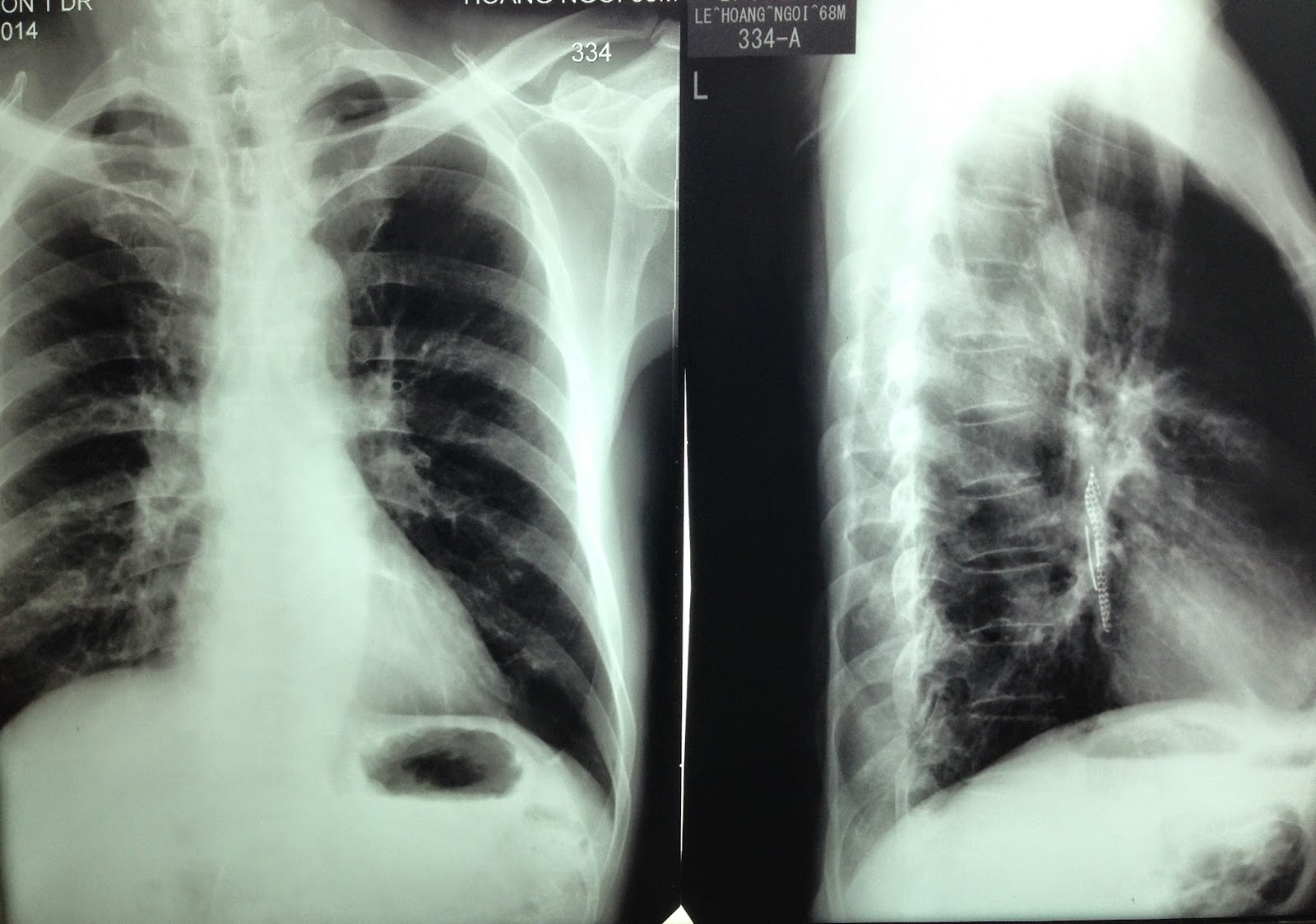
Chest XRay on PA was normal, but on LATERAL VIEW there was something in retrocardiac space (chest XRay).
MSCT scan detected foreign body in the midlle of his esophagus ( see 4 CT pictures).
↧
CASE 251: PAIN at RIGHT HIP, Dr PHAN THANH HẢI, MEDIC MEDICAL CENTER, HCMC, VIETNAM
Man 52 yo with history of pain at his right hip joint 2 years prior, recently the pain is getting more severe, cannot walking (photo).
Plain XRay film of the pelvis looked like normal right and left hip joints.
Upon ultrasound the right hip joint showed widering of the hip joint space with fluid collection, and abnormal echostructure of the head of femoral bone (see 3 ultrasound pictures at right hip).
Ultrasound examination of the right hip report was abnormal in suggesting arthrosis of right hip joint).
MRI of the hip joint showed that right femoral head in necrosis and hydarthrosis, and small change also at left hip joint.
↧
↧
CASE 252: NECK PAIN in LEMIERRE'S SYNDROME, Dr PHAN THANH HẢI, MEDIC MEDICAL CENTER, HCMC, VIETNAM.
Girl 13 yo, for one week sore throat and fever, being treated with antibiotics, now pain at right neck.
Ultrasound at the neck detected on right side some lymph nodes 2-3 cm at posterior SCM. And internal jugular vein dilated, big diameter 2, 2 cm black lumen, no flow, cannot compressible (see US pictures of CCA and IJV on right side).
Ultrasound at the neck detected on right side some lymph nodes 2-3 cm at posterior SCM. And internal jugular vein dilated, big diameter 2, 2 cm black lumen, no flow, cannot compressible (see US pictures of CCA and IJV on right side).
Meanwhile, on the left neck, there were normal flow of CCA and IJV ( see video clip).
CDI OF CCA AND IJV on R NECK
VIDEO 2: CROSSECTIONAL
What is your emergent thingking? What is the lab you must execute evaluation ?.
Blood tests: WBC= 15k.. neutro 40%, CRP=20mg/l very high, D-Dimer=500ng/ml Bacteriology bloodculture is on the way..
Based on CLINICAL INFECTION.and LAB REPORTS, and ULTRASOUND IMAGES.of THROMBOSIS OF IJV, suggustion LEMIERRE SYNDROME.
Urgent treatment with IV ANTIBIOTICS and ANTICOAGULATION DRUGS.
REF..HISTORY OF PROF ANDRE ALFRED LEMIERRE.
↧
CASE 253: GOSSYPIBOMA (TEXTILOMA) POST CAESAREAN SECTION for a YEAR, Pham Hong Dong,M.D; Nguyen Duc Duy Linh,M.D; Phu Van Tuot,M.D; Nguyen Ngoc Xuan Giang,M.D., MEDIC Binh An Kien Giang Hospital
A 26 year-old female patient who had complained mild pain at her pubic region presented lower abdominal pain a month prior. She overwent a caesarean section a year ago for delivery her child.
Ultrasound findings:A cystic mass (about 83x46 mm) containing distinct internal hyperechoic wavy, striped structures.
CT Scan abdomen: A mass of 11 x 9 cm with thicken enhancing walls was seen in pelvis.
But diagnosis of gossypiboma was made and at laparotomy: a surgical sponge (18x22 cm) with adjacent inflammatory tissue and pus were removed successfully.
DISCUSSION:
A diagnosis of gossipiboma pre-op seems to be very difficult that always need skill and experience. Because of imaging findings of gossypiboma are nonspecific and complexe so the right diagnosis in pre-op is still acchived about 1/3 of cases in literature.
But whenever an unknown mass into abdomen with exist surgical scare that should dissolve it may be a gossypiboma or not.
↧
CASE 254: CASTLEMAN DISEASE in COLONIC MESENTERY, Dr JASMINE THANH XUÂN, Dr PHAN THANH HẢI, MEDIC MEDICAL CENTER, HCMC, VIETNAM
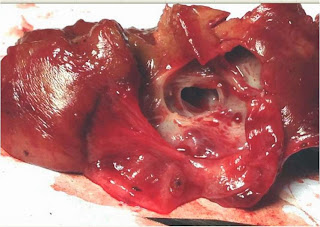
A 22 yo female patient with a mass of right abdomen which was detected by ultrasound check-up and thought to be a mesenteric tumor or a lymph node in mesentery. It was well-bordered and vascular structure without any symptom.
MSCT confirmed the 14x17mm mesenteric tumor in right abdomen with CE enhancement.
Open surgery removed the mass from posterior space of right colonic mesentery.
Microscopic result is a Castleman disease in mesentery, whichis an uncommon lymphoproliferative disorder that may be localized to a single lymph node (unicentric) or occur systemically (multicentric).
It was a second case at Medic Center.
The first case of Castleman disease was posted in 2010.
CASTLEMAN DISEASE in RETROPERITONEAL SPACE at MEDIC CENTER
↧
CASE 255: PERI-BREAST TUMOR, Dr PHAN THANH HẢI-Dr JASMINE THANH XUÂN, MEDIC MEDICAL CENTER, HCMC, VIETNAM
Woman 34yo, in palpation detected herself at RUEQ one mass suspected breast tumor.
Mammography confirmed one mass with macrocalcification.. at 1h site of right breast with dense tissue (see 2 mammo pictures).
Ultrasound scanning at right breast detected one hypoechoic ellypsoid mass with size of 3cmx2cm in major pectoralis muscle, Upon CDI scan this mass was hypovascular, and elastoscan was hard tissue, no axillary lymph node ( see ultrasound scan B mode, CDI, elasto).
What is your suggestion of diagnosis ?.
MRI of mammary glands were done, this mass was retromammary, inside major pectoralis muscle on right site. The signal suggestion was hemangioma (see 2 MRI pictures).
FNAC report was compatible with retromammary hemangioma.
Operation removed completly this mass; microscopic report was cavernous hemangioma.
REF case pdf.
↧
↧
CASE 256: LIVER FUNGAL INFECTION in HIV-INFECTED PATIENT, Dr LÊ ĐÌNH VĨNH PHÚC, Dr VÕ NGUYỄN THÀNH NHÂN, MEDIC MEDICAL CENTER HCMC, VIETNAM
A 30 year-old married woman, suffered from weight loss, fatigue, not fever, not abdominal pain. She has scanned by abdominal ultrasound at a province hospital detecting multifocal lesions in liver. Her doctor thought her liver hemangioma.
At MEDIC center, ultrasound scanning detected multi-hyperechoic masses with regular border, no vascular proliferative, no around liver parenchyma edema, no necrosis fluid, size of 0.5 to 2cm in right and left lobe.
CT Scan of liver was done with many reduced density lesions in the right and left lobe. The lesions were slight contrast enhancement. Some lesions were higher than in the center area.
Blood test with WBC normal, transaminases slight increase, HBsAg negative, anti-HCV negative. The important noticeable result is thatanti-HIV positive (ELISA).
The findings of ultrasound, CT Scan and blood test suggested liver fungal infection in HIV-infected patient. This patient was treated with anti-fungal drugs. Fungal infection is a common opportunistic disease in HIV-infected patient. Among the fungal opportunistic infections, Coccidioides immitis and Histoplasma capsulatum are those most likely to involve the liver [1]. Fungalliverabscessdiagnosisremains achallenge fordiagnostic imagingandclinical.
What is your suggestion of diagnosis?
References:
1. Anthony S. Fauci; H. Clifford Lane (2010). “Human immunodeficiency virus disease: AIDS and related disorders”. Harrison’s infectious diseases. Mc Graw Hill. p. 847
↧
CASE 257: KNEE PAIN, Dr PHAN THANH HẢI, MEDIC MEDICAL CENTER, HCMC, VIETNAM
Man 35 yo, 3 days ago, pain at right knee cannot move, fever, no history of trauma. Clinical examination of right knee: hot and swelling at suprapatellar area (see photo).
Ultrasound first scanned at the right knee, detecting swelling of the suprapatella recessus with homogeneous fluid (2 ultrasound pictures).
Blood test confirmed this infectious status with rising WBC and CRP.
For make sure the diagnosis: puncture of the knee joint with ultrasound guided..removing the yellowish synovial fluid.
Diagnosis of this case is acute bacteria infection of the knee joint, emergency treated with antibotic and analgesic drugs.
DISCUSSION: in acute case ultrasound guided puncture of the joint is fast action for fast diagnosis.
↧
CASE 258: Multiple Intraabdomen Nodules, Dr PHAN THANH HẢI, MEDIC MEDICAL CENTER, HCMC, VIETNAM
Man 54 yo, 2 years after operation.for acute obstruction of small bowel by tumor of intestine, unknown microscopic report.
Now he had pain in the pelvis. (photo of the skin scar operation).
Ultrasound report were multiple nodules in pelvis looked like grappe fruit
adherence over urinary bladder wall (2 ultrasound pictures).
MSCT CE of abdomen detected multiple intramesenteric round tumors looked like lymph nodes.
All blood test and cancer markers were normal.
↧
CASE 259: PLEURAL EFFUSION, Dr PHAN THANH HẢI, MEDIC MEDICAL CENTER, HCMC, VIETNAM
Woman 23 yo for one month cough and dyspnea, no fever.
Ultrasound of the thorax detected left pleural effusion with collapsed lung and one mass covered anterior mediastinum to external pericardium (see 3 ultrasound pictures of the left lung).
MSCT with CE of the chest confirmed a big mediastinal tumor with pleural effusion which displaced the heart to the right side (see 3 CT pictures).
↧
↧
CASE 260: ODDI TUMOR, Dr PHAN THANH HẢI, MEDIC MEDICAL CENTER, HCMC, VIETNAM
Man 40 yo pain at RUQ with dark urine.
Ultrasound first detected big gallbladder with very think wall, no stone, CBD in dilatation just to end of CBD, no tumor of pancreas (2 ultrasound images).
MSCT CE detected dilated biliary system intra and extrahepatic ( 3 CT images).
Blood tests, CEA, CA-19-9 were normal.
What is your suggestion for diagnosis and your next step ?
ERCP not successful.
Biopsy of the tumor but the microscopic result was negative.
Ooen operation for exploration, surgeon detected a hard massat the head of pancreas.
Whipple operation was performed (specimen of tumor of Oddi).
Microscopic report was adenocarcinoma ( patho images)
↧
CASE 261: HARD BREAST like AMBARELLA FRUIT, Dr PHAN THANH HẢI, MEDIC MEDICAL CENTER, HCMC, VIETNAM
Woman 47 yo, she herself detected at the right breast one mass, slow growth, painless like one AMBARELLA fruit.
Ultrasound first detected this mass in size of 4 cm, at 10 am clock at the right breast, with many white spots as calcification. Color Doppler finding also was a hypovascular mass.
Using elastoultrasound, the mass was very hard, scale blue green color on elastogram, no detected axillary lymph nodes.
Mammography also detected mass and microcalcification.
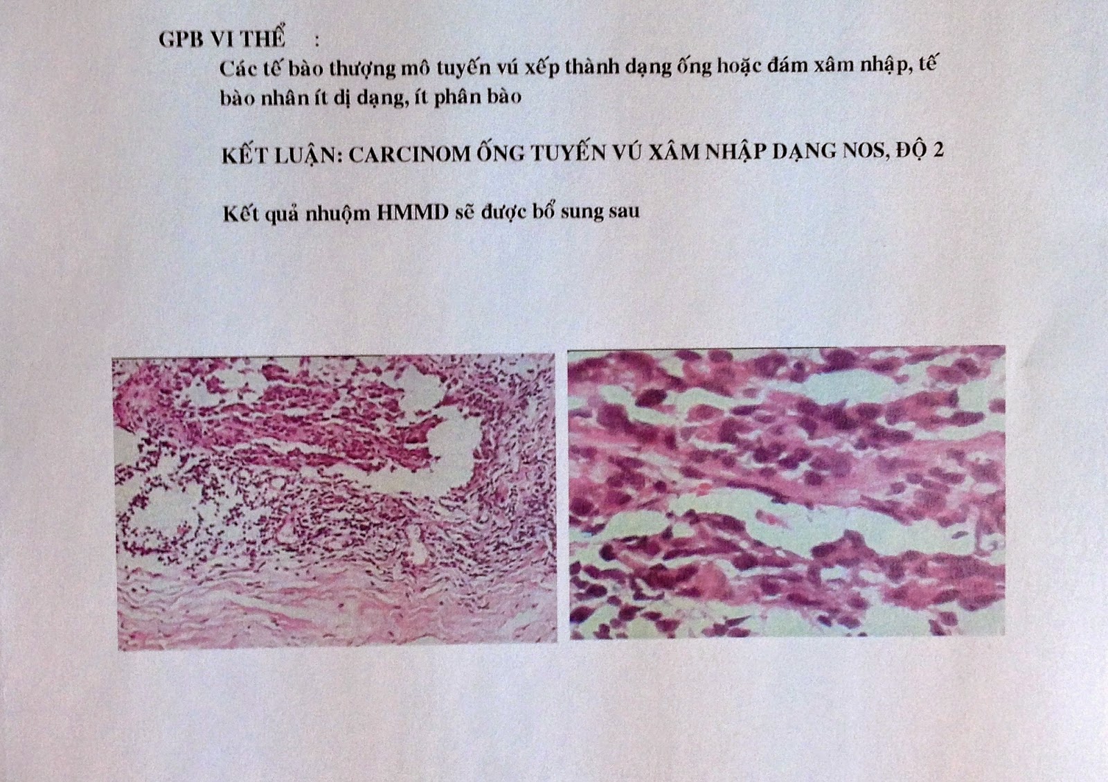
Core biopsy with ultrasound guided reports microscopic INTRADUCTAL CARCINOMA, STAGING T2N0MX.
SUMMARY: BREAST CANCER IS EASY DIAGNOSED BY ULTRASOUND WITH ELASTOSCAN of THIS VERY HARD TUMOR LIKE GREEN AMBARELLA FRUIT.
↧
CASE 262: BIG ABDOMEN, Dr PHAN THANH HẢI, MEDIC MEDICAL CENTER, HCMC, VIETNAM
Man 50 yo, one week ago, onset periumbilical pain and abdominal distension, no defecation nor fever.
Chest Xray, and abdomen standing plain film showed the water-air level in intestine, suggesting bowel obstruction.
Ultrasound found out colon dilatation, filling water and moving circular around with hyperperistalsis (see video).
MSCT of abdomen in emergency detected dilated right colon and small intestine, retroperitoneum edema arround the pancreas and radiologist suggested that pancreatitis.
Blood test: WBC rising 12k, amylasemia normal.
Operation laparotomy detected all bowel in dilatation but no necrosis, no tumor obstruction.
Many white spots like candle intra peritoneum.
Retroperitoneal space edema. Surgeon said chronic pancreatitis.
Operation laparotomy detected all bowel in dilatation but no necrosis, no tumor obstruction.
Many white spots like candle intra peritoneum.
Retroperitoneal space edema. Surgeon said chronic pancreatitis.
Discussion of this case: clinical findings were abdominal pain and distension for one week. XRay and ultrasound found out bowel obstruction and CT detected pancreatitis, but blood test amylasemia was 17 unit.
Surgeon decided operation by bowel obstruction.
Now report is chronic pancreatitis, it is a rare case with normal amylasemia in acute pancreatitis.
Now report is chronic pancreatitis, it is a rare case with normal amylasemia in acute pancreatitis.
REFERENCE: case report
↧
CASE 263: BUFFALO'S NECK, Dr PHAN THANH HẢI, MEDIC MEDICAL CENTER, HCMC, VIETNAM
Man 27 yo, dental pain on right mandibular for one week, he detected the right side of neck along SCM muscle getting hot and swelling, pain and fever (see photo).
He was treated with antibiotics and went to ultrasound scanning. Sonologist detected this neck mass beeing like abscess by fluid collection on right and left neck (see 3 photo and video).
MSCT found out the right mass along SCM muscle and one other mass on left side nearby thyroid gland.
Puncture this mass removed the pus but direct examination with gram stain no bacteria.
Operation for drainage.
↧
↧
CASE 264: ASCITES, Dr PHAN THANH HẢI, Dr LÝ VĂN PHÁI, MEDIC MEDICAL CENTER, HCMC, VIETNAM
Woman 31yo 3 days ago epigastric pain.
She came to MEDIC for gastroscopy and result was gastritis, but ultrasound of abdomen detected free fluid of ascites around border of liver and small stone in gallbladder (see ultrasound images).
Liver was normal, and ultrasound at pelvis detected one mass on the left side of uterus, round, 5cm diameter, solid mass with small vessel inside and RI low ( see ultrasound images). Sonologist susgested an ovary tumor in rupture.
MSCT with CE of this mass on left lateral of uterus...with CE enhance like a nidus of pregnancy in rupture with a lot of blood clots in abdomen ( see 3 CT pictures).
Blood tests : CA-125 rising 125 UI/mL and betaHCG rising 134UI/mL
WBC 15K with neutro 75%, Hct 29%.
Emergency operation in BINH DAN hospital detected hemoperitoneum due to rupture of tubal pregnancy (photo).
DISCUSSION:
Epigastric pain is a common indication to gastroscopy that was not available for this case.
ULTRASOUND of ABDOMEN MUST BE FIRST CHOICE for CASE.
BLOOD TEST CA-125 RISING NO MEANING TO OVARY CANCER.
Beta HCG was most sensitive for diagnosis of this case.
↧
CASE 265: FISH BONE IN GALLBLADDER: Dr PHAN THANH HẢI, MEDIC MEDICAL CENTER, HCMC, VIETNAM
Man 49 yo, pain in RUQ one week ago like gastric ulcer.
Ultrasound of abdomen suggested gastric cancer invasive to gallbladder and liver.
Gastroscopy and biopsy ruled out gastric cancer.
MSCT with CE detected abscess due to perforated fundus of gallbladder and one foreign body like a fish bone, 3cm in length, intra gallbladder (see 3 CT pictures).
Ultrasound of abdomen again for verify diagnosis.also made same information which was abscess due to fish bone penetrated through gallbladder wall to liver border.
(see 2 ultrasound images and video clip).
Blood tests were normal.
↧
CASE 266: COLO-COLIC INTUSSUSCEPTION, Dr PHAN THANH HẢI, MEDIC MEDICAL CENTER, HCMC, VIETNAM
Man 38 yo, epigastric pain crisis on periodic treatment like gastritis but not response.
Ultrasound first in emergency at MEDIC detected one mass near gallbladder with multilayer cover as OINION SIGN, and a central cyst.
This mass was in transverse colon. Sonologist suggested a colocolic intussusception (see 04 ultrasound images and video clip).
Do you have any idea about the cyst in an intussusception mass?.
MSCT with CE showed this mass in transverse colon with cystic mass looked like appendicular mucocele.
↧


.jpg)
.jpg)
.jpg)


