Quantcast
Channel: VIETNAMESE MEDIC ULTRASOUND
Viewing all 624 articles
Browse latest View live

CASE 228: Thyroid Toxicosis Periodic Paralysis, Dr PHAN THANH HẢI. MEDIC MEDICAL CENTER, HCMC, VIETNAM

$
0
0

Thyroid Toxicosis Periodic  Paralysis
Man 47 yo, 3 times  paralysis  at  get up in the  morning, he  came to hospital  for  emergency  perfusion  potassium and to  MEDIC  for check- up.
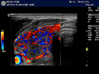
Ultrasound  of abdomen  no abnormal  detected,  no adrenal tumor, but  ultrasound  of thyroid gland is black , CDI hypervasular , typical of  hyperthyroidis .and  soft  with  elastoscan.


Blood test are very low TSH  , high T3, T4, TPO.
EMG wassignalof  hypokalemia.


Thiscase isTpP. Medical treatmentof anti thyroidism  is requested.

REFERENCES:


CASE 229: UMBILICAL TUMOR, Dr PHAN THANH HẢI, MEDIC MEDICAL CENTER, HCMC, VIETNAM

$
0
0
Girl 7 yo detected umbilical foramen distention many years ago {see photo}. No clinical symptome.
On utrasound scanning it was well - bordered cystic tumor with septation. Color Doppler showed small vascularized intraseptal tumor(2 images).
MSCT with CE, tumor was not related with intra abdomen; its structure was very high contrast enhanced in late phase.( 3 images)
This case had been examined by many doctors. At first one sonologist suggested umbilical hernia, second senologist diagnosed as lymphangioma, another sonologist said hemo-lymphangioma. One radiologist said as dermoid cyst or fibroma. In operation removing this mass (see photos).

CASE 230:CONTRALATERAL BREAST CANCER (CBC), Dr PHAN THANH HẢI, MEDIC MEDICAL CENTER, HCMC, VIETNAM

$
0
0
WOMAN 64 YO, 10 YEARS BEFORE SHE WENT THROUGH RIGHT MASTECTOMY BY BREAT CANCER [ T2N1MX ]. HISTOLOGY REPORT POST OP WAS DUCTAL CARCINOMA , NOS, ER, PR NEGATIVE. NO CHEMO NOR RADIOTHERAPY.

NOW SHE DETECTED BY HERSELF ONE MASS AT LEFT BREAST. ON ULTRASOUND IT WAS A TUMOR WITH CALCIFICATION, SIZE OF 4 CM AND BIG AXILLARY NODE (2 IMAGES).


MAMMOGRAPHY ALSO SUSPECTED MALIGNANT TUMOR.

MSCT OF THORAX : NO RECURRENT ON RIGHT SIDE, THE LEFT BREAST HAD TUMOR NEAR THE NIPPLE AND AXILLARY NODE. NOTHING DETECTED ANOTHER LESION OF OTHER ORGAN.


WHAT IS YOUR IDEA ? IT IS METASTASIS TO LEFT BREAST FROM THE FIRST BREAST CANCER OR SECOND CONTRALATERAL BREAST CANCER?

CASE 231: INTRALIVER TUMOR, Dr PHAN THANH HẢI, MEDIC MEDICAL CENTER, HCMC, VIETNAM

$
0
0

Woman 36 yo with long history of acute hypertension crisis.

Ultrasound of abdomen for check-up detected one mass at left lobe  of liver,  size of 11 cm-8,5 cm which had  round border  with central mass being thought  a scar necrosis( 2 pictures). Sonologist suggested a FNH or HCC.


Blood tests are normal AFP,  not infected HBV ,or HCV.

MSCT with CE of this tumor was well–bordered, blood supplying from liver tissue around, hypervascular with rapid washed-out contrast. This tumor had displaced liver vein. Radiologist diagnosis was a liver tumor as HCC.


Ultrasound guided biopsy was reported that a HCC.

Operation removed this tumor completely and it was not a liver tumor, but was one tumor outside liver .

Microscopic report was malignant pheochromocytoma.

Do you review if  those pictures  are correct with the end report or not?

CASE 232: SORE THROAT AFTER COFFEE DRINKING, Dr PHAN THANH HẢI-Dr TRẦM THỊ TÚ HƯƠNG, MEDIC MEDICAL CENTER, HCMC, VIETNAM

$
0
0
Woman 50 yo more severe sore thoat after drinking black coffee cup, then she came to ENT hospital for endoscopy, CT of the neck: nothing detection while the neck was swelling, painful and fever. It is LEMIERRE's syndrome.


 MEDIC ultrasound of the neck detected on left lobe of thyroid one echo rich line, long of 3 cm, in an abscess and another abscess on right lobe of thyroid also.


Sonologist said it was a toothpick penetrating to the left thyroid gland.

 
WBC rising of 15k with 87% neutro and CRP was  high : 36mg/L.

MRI at the neck was done and detected a black line as the toothpick.


Emergency endoscopy detected the stuck toothpick in the throat and removing it out (see photo).


 This patient was treated with antibiotic and nasogastric feeding.

REFERENCES:


CASE 213: BILOBED GALLBLADDER, Dr PHAN THANH HẢI, MEDIC MEDICAL CENTER, HCMC, VIETNAM

$
0
0

Woman29 yo,  recurrent pain atsubhepatic area.
Ultrasound detectedstone in gallblader with thickening wall and one cystic mass withsize of 2 cm  nearby. Sonologist reported that wasCBD cystandchroniccholecystitisdue to stone [3 images].


MSCT  also had samereport as ultrasound report (see  image CT).


Surgeonordered  MRCP. The MRI report was different which thought about  bilobar gallbladder that had 2 parts: one part had stone and  thickeningof the wall, and another part wasconnected to the neckof gall bladder which had  normalwalland normal CBD.


Endoscopic surgeryforremoving gall bladderwithstone in its fundus.
Macroscopic specimen of this gallbladder wascorrelated with 3 modalities of imaging: ultrasound, CT and MRI.


 REFERENCE


CASE 233: ABDOMINAL MASS AFTER TRAUMA, Dr PHAN THANH HẢI - Dr PHÙ VĂN TUỐT, BÌNH AN HOSPITAL, VIETNAM

$
0
0
MAN 23 YO, AFTER ONE WEEK UNDERWENT A BLUNT TRAUMA NEAR PARAUMBILIC AREA BY BICYCLE ACCIDENT, HE WAS GETTING WORSE MORE AND MORE  BY VOMITTING, AND CANNOT EATING. 

CLINICAL EXAMINATION OF ABDOMEN REVEALED A MASS OVER RIGHT KIDNEY, POSITIVE REBOUND TENDENESS ( PHOTO).


ULTRASOUND FIRST SHOWED THE STOMACH WAS DILATED. THE  MASS WAS AT DUODENUM D2-D3, SIZE OF 5 CM, CYSTIC STRUCTURE BUT NO FREE FLUID IN HIS ABDOMEN ( 3 U/S IMAGES).


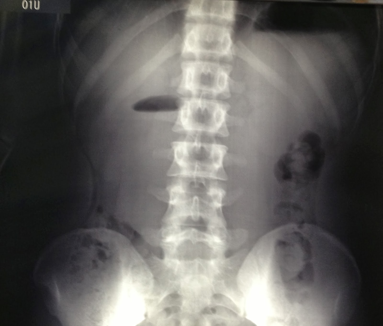
XRAY of ABDOMEN in STANDING  and BARIUM MEAL  SHOWED THAT DOUDENUM  in OBSTRUCTION.


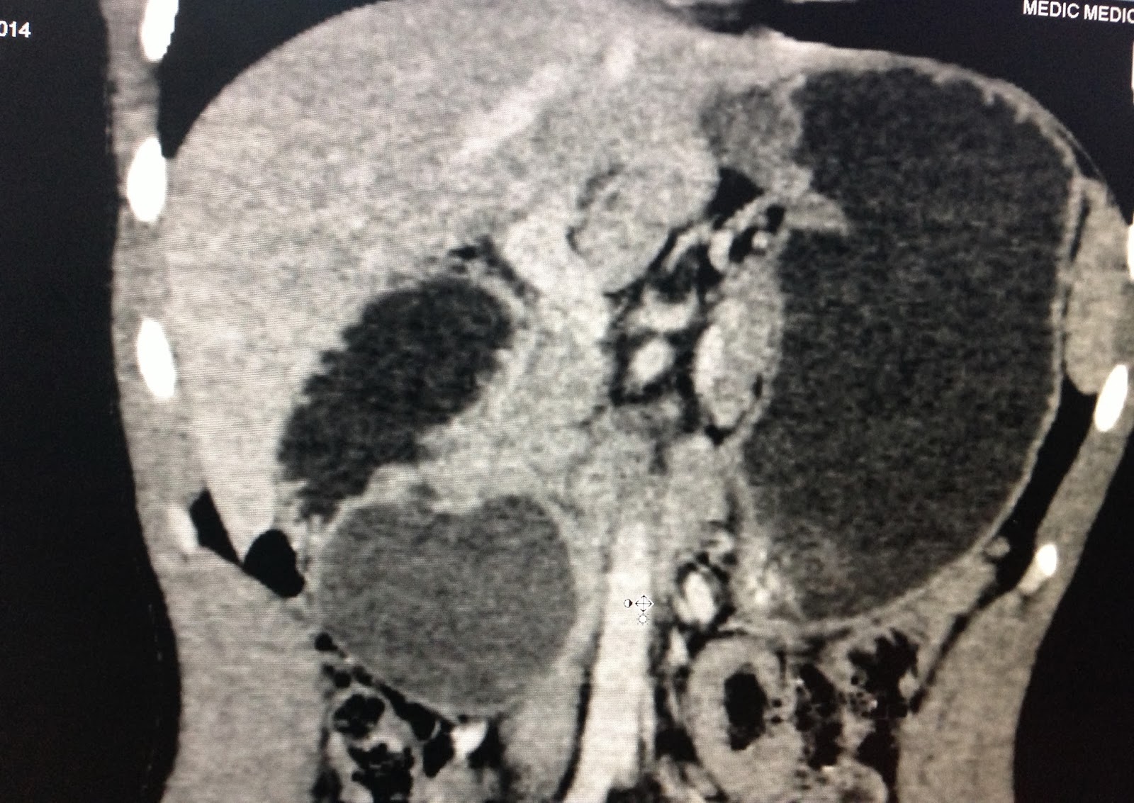
MSCT of abdomen: this mass was from duodenum wall,  cystic structure,  size of 5 cm. Radiologist made diagnosis  of hematoma intraduodenum wall without bleeding into abdomen.


Medical treatment by nasogatric suction  and  wait and see the progress.
 
AFTER 4 DAYS for  NASOGASTRIC SUCTION and IV FLUID REPLACEMENT, NASO GASTRIC TUBE HAD BEEN REMOVED and XRAY BARIUM MEAL for  BEING SURE NO OBSTRUCTION of  DUODENUM. HE CAN DRINK and  EAT  and  RECOVERY STAGE DURING SO FAST (see XRay barium meal on  Jan 24, 2014).
IT IS FIRST STEP of  SUCCESS of MEDICAL TREATMENT for HEMATOMA DUODENAL WALL POST TRAUMA.

REFERENCES:


CASE 234: A LATERAL MASS of the NECK, Dr PHAN THANH HẢI, MEDIC MEDICAL CENTER, HCMC, VIETNAM

$
0
0


Man 56 yo for a long time being follow up with thyroid nodule, now he detected the palpable mass at right neck,  situation at middle SCM .
Ultrasound suspected lymph node with B mode ultrasound ( picture 1,2: thyroid nodule) on right side of the neck.


For ruling out a thyroid cancer,  a FNAC was performed the thyroid mass and the result was colloidal goiter.


MRI of the neck also suspected this mass between carotid internal and external artries ( 2 MRI pictures).


For diagnosing this neck mass we did CDI for guiding FNAC, but this mass was very high vasculature and situated between ICA and ECA. (see 2 CDI images  of cross section and longitudinal scanning this mass).


What is your proposal for another step of diagnosis?


CASE 235: PERIAORTIC MASS, Dr PHAN THANH HẢI, MEDIC MEDICAL CENTER, HCMC VIETNAM

$
0
0
Man 63 yo,  medium arterial hypertension with umbilical battement with the heart beat.

Ultrasound of abdomen detected  kidney on 2 sides  in hydronephrosis due to ureteral compression by the mass covering arround the abdominal aorta. Of which structure was echo-poor looked like a cyst. The aorta was pull up near the umbilicus (see 5 ultrasound pictures ).


MSCT with CE (4 CT pictures); CT 1..kidney hydronephrosis, CT 2, CT 3, CT 4 .. intratumor abdominal aorta.


Blood tests were normal WBC, PSA, CEA, with beta MICROGLOBULIN was 2298ng/ml (N <2164

Urologist  suggestion was retroaortic lymphoma and he put 2 jj stent intra ureter and operation for biopsy this mass.
Open operation detected this mass being fixed and covering aorta and compressing completely causing collaped  IVC.( see foto per op.1 and 2..right ureter with jj stent inside).


Wait for pathology report.

CASE 236: LYMPHOMA MIMICKING PANCREATIC TUMOR and METASTASIZING LYMPH NODES in ABDOMEN and NECK, Dr LÊ THANH LIÊM - Dr LÝ VĂN PHÁI, MEDIC MEDICAL CENTER, HCMC, VIETNAM

$
0
0
       58yo  male patient, slight fever  and  fatigue for 6 months, a lymph node on right neck getting bigger.  
           Lymph node at supraclavicular area was hypervascular and looked-like a cyst on color Doppler ultrasound. 

      Ultrasound  of abdomen detected epigastric tumor between left lobe of liver and pancreas, hypervascular and celiac vessels penetrating the tumor. Some nodes were  around the tumor, and body of pancreas not well-bordered. 


     Ultrasonologist thought about lymphoma infiltrating in abdominal cavity and neck,  but keepng a different diagnosis with pancreatic tumor metastasizing abdominal and neck lymph nodes.
       MSCT confirmed lymph nodes lymphoma in abdomen and no pancreas tumor but lower density of the body.


     FNAC of neck lymph node and the result was big B cell lymphoma. So this is another case of big B cell lymphoma with the evident of neck FNAC result.


CASE 237: HORSESHOE TUMOR, Dr PHAN THANH HẢI - Dr NGUYỄN TUẤN VINH, MEDIC MEDICAL CENTER, HCMC, VIETNAM

$
0
0

Man 57 yo, diabetes and  gallbladder stone. He's got Murphy's pain.
Abdominal CT with CE detected the  horseshoe kidney with tumor of 3cm.( Fig.CT 1).

I

Ultrasound  for  evaluation of this tumor  was hypoechoic, at the  left part of  horseshoe kidney and hypovascular( Fig. US 1, US 2).


MSCT 640 slices performed with CE again for staging  of this tumor (see 4 CT pictures).


One urologist  said no biopsy.
Operation  for  removing left part of  horseshoe kidney.

Macroscopic appearance of  tumor  is well-bordered into parenchyma of kidney.  Microscopic report is clear cell carcinoma.

REFERENCE:


CASE 238: HBV PATIENT with 2 SIDE KIDNEY TUMORS, Dr LÊ THANH LIÊM, MEDIC MEDICAL CENTER, HCMC, VIETNAM.

$
0
0

  Male patient 34yo from  Binh duong province in follow-up  from HBV infection since 2006. He had got 2 kidney cysts and nothing abnormal, but now B-mode ultrasound detected kidney tumors on his 2  sides. Both 2 kidney tumors hyperechoic, homogenous seem to be looked like AML tumor. But they were different on color Doppler, hypervascular, with venous and arterial patterns, maybe oncocytoma or RCC (renal cell carcinoma).
On 3 D ultrasound reconstruction  the left kidney tumor more clear than on B-mode ultrasound.
And elastographic ultrasound (ARFI) proved tumors  more hard than kidney parenchyma.

 
CT proved 2 kidney tumors on 2 sides of one patient.


He underwent 2 operations for removing 2 kidney tumors, on left side first, partial nephrectomy, with microscopic result of papillary RCC, and right one, total nephrectomy for pRCC again after half of month.
 
pRCC is a unusual kidney tumor, mimicking AML on B-mode ultrasound so we should  use color Doppler to examine kidney tumors.

REFERENCE:


CASE 239: COLON TUMOR and WALLED-OFF ABSCESS, Dr PHAN THANH HẢI, MEDIC MEDICAL CENTER, HCMC, VIETNAM

$
0
0

Man  dob 1951,  too painful  in left  flancwith  digestive trouble  in  defecation.
Ultrasound  first   detected  a hypoechoic  mass   along   left  flanc with  pseudocervix sign. Sonologist   suggested that  descending colon tumor and  peritumoral  abscess ( see3  ultrasound pictures).


MSCT  with CE   diagnoses this mass  being  colon tumor  subocclusion and  abscess   arround tumor ( see CT  pictures).


Colo-endoscopy  detected     lumen stenosis of   descending tumor, but  biopsy  this  tumor  2 times   all  report  were free of cancer cell.


Blood test   WBC  not  rising , CEA, CA 19-9  were negative.
Open operation was done in  detecting  big mass  due to colon cancer  invading to around  structures,   .. making   the wall-off mass ( see  foto).


Wait  for microscopic report.

CASE 240: ADRENAL GLAND TUMOR SECRETING ALDOSTERONE, Dr LÝ VĂN PHÁI, MEDIC MEDICAL CENTER, HCMC, VIETNAM

$
0
0


Female patient 22 yo from Daklak province, student. Arterial hypertension for 1 year and weknesss of 4 limbs in effort and remaining normal in rest.
Abdominal ultrasound detected  right adrenal mass of 21x28x24mm, well bordered which was thougt to be an adrenal tumor.


MDCT confirmed an adrenal tumor on right side.


Blood tests:  Kalemia =1.77mmol/l ( N= 3,5-5), Aldosterone  = 380pg/ml ( N= 10-210).

Operation was done and microscopic report was a benign adrenal tumor.


REFERENCES:
http://en.wikipedia.org/wiki/Primary_aldosteronism
http://www.studyblue.com/notes/note/n/en1-03-adrenal-glands-1/deck/1600308

CASE 241: SEMINAL TUBERCULOSIS : Dr NGUYỄN MINH THIỀN, Dr PHAN THANH HẢI, MEDIC MEDICAL CENTER, HCM C,VIETNAM

$
0
0

Man  51 yo, one month ago suffered from lowgrade  fever and dysuria. Chest Xray was in suspection of  lung tuberculosis.


Abdomen ultrasound  detected  many  lymph nodes around  aorta ( 2 ultrasound pictures) scanning  at level  of pancreas  (cros-sectional and longitudinal scan).


Ultrasound at pelvis detected one hypoechoic mass of right seminal vesicle, no vascular structure intra this mass.


MSCT also detected one mass on right site of prostate, suspected  abscess (2 CT pictures).


TRUS guided puncture this mass withdrew out the pus in white color  looked like caseum. Analysis this pus was  positive PCR tuberculosis.


Conclusion of this case :  tuberculosis of the lung, lymph nodes, and  seminal  vesicular mass.  Medical treatment  of antituberculosis drugs was setting up for the patient.

REFERENCES:


CASE 242: IVC TUMOR, Dr PHAN THANH HẢI, Dr LÝ VĂN PHÁI, MEDIC MEDICAL CENTER, HCMC, VIETNAM

$
0
0


Woman 35 yo, pain at epigastric area. Ultrasound in a provencial hospital suspected liver tumor, and  reviewing of ultrasound at MEDIC center.
This mass was hypoechoic structure with  size of 7cm X 5cm along of upper portion of IVC in covering over right kidney and duodenum (see 4 ultrasound pictures). 


MSCT with CE of  this mass was slow enhancement, and in invasion of the wall of IVC ( see 3 CT pictures).


Gastroendoscopy was ruling out a tumor from duodenum.
Blood test : CA19-9 rising of 62.58 UI/mL
What is your suggestion of diagnosis and planning for treatment ?.

Discussion: this case did not have GI tract  symptoms , no endocrinal effect, and the mass  situated  in retroperitoneum  and  IVC compression, we can rule out  liver tumor, GIST of duodenum, kidney tumor. The rest of retroperitoneal tumors  near I V C are  neuroendocrine tumor  or leiomyosarcoma, liposarcoma with CA 19-9 rising, we cannot explain  what is  situation. Pre-op  diagnosis is retro peritoneum tumor looked like sarcoma. Operation was done for removing completely  this mass  with one part of IVC (see 2 operation samples). Microscopy report was a retroperitoneal leiomyosarcoma.


REFERENCE:


CASE 243: GALLBLADDER TUMOR, Dr PHAN THANH HAI, MEDIC MEDICAL CENTER, HCMC, VIETNAM

$
0
0
Man 69 yo,  pain  at  epigastric area, no  fever,  no GI tract trouble.
Abdominal  ultrasound  detected  abnormal  gallbladder: thickening of the wall with one  mass  at gall bladder  fundus  invasion to liver, suggestion of gallbladder abscess ( see 4 ultrasound pictures).


Blood test  are normal:   WBC  not rising, CA 19-9  normal
MRI with CE gado  showed this mass  in  high  enhancement  invasion in to  liver and  transverse colon, the  biliary system was  normal.


With  this clinical situation and blood tests, US first and  MRI,  what is your diagnosis ?

DISCUSSION: this case  had no  clinical signs of acute  cholecystisis, no stone  in gallbladder; ultrasound  showed  the  wall of  the gallbladder   perforated and  adherent , invading  liver  tissue, this mass was  hypovascular  in protruding  into lumen of gallbladder as a tumor and  going to extra wall of gallbladder.

Open operation  with  diagnosis  of  tumor of gall bladder (surgeon  removed  gallbladder  and  resection one part of liver  and  great omentum).  Specimen was  hard and necrosis.


Microscopic with imunohistostaining is leiomyosarcoma of the gallbladder..it is very rare  case in the word  had been published.
REFERENCE:


CASE 244: AVM and RETROPERITONEAL MASS, Dr PHAN THANH HAI, DR LY VAN PHAI, MEDIC MEDICAL CENTER, HCMC, VIETNAM

$
0
0


Man 31 yo, one week ago complained  of epigastric pain and vomiting.
Clinical examination he had red skin on the right thorax and atrophied muscles of right arm.


Emergent abdominal ultrasound scan showed  one mass of 4cm located near the head of pancreas, at processus uncinatus which  compressed duodenum..
(see 3 ultrasound pictures.. P1.color doppler at right subclavicule suspected A-V-M) , P4 .P5..cross scan and long scan this mass at the head of pancreas.)


Gastroendoscopy went down just to duodenum but nothing detected.
MSCT with CE: this mass was in retroperitoneum compressing duodenum D2,
contrast injection was slowly enhancement , but it had air in the  mass ( see 4 CT  with CE pictures CT1, is angiogram of right axillary artery, CT2. this mass with air inside, CT3, relation with right kidney and aorta, CT4 vascular SMA and mass).


Blood tests were no abnormal.
What is your suggestion for diagnosis ?.

CASE 245: CERVICAL VAGUS NERVE SCHWANNOMA, Dr PHAN THANH HAI - Dr LAM CAM TU, MEDIC MEDICAL CENTER, HCMC, VIETNAM

$
0
0


Woman 51yo detected lateral left neck mass, no pain (see foto ).


ENT doctor did endoscopy for rule out a cavum cancer, and FNAC did not detect cancer cell metastasis to this mass.


Ultrasound report was an ellipse mass of 3 x 5 cm,  well-bordered,  hypoechoic, hypovascular with small cystic formation which deplaced the CCA and internal jugular vein (IJV) that well confirmed by 3D vascular CT angio (see 4 CT pictures).


Discussion:  This 51 yo woman with the mass in upper portion of left lateral neck, painless for a long time suggesting malignancy.
Ultrasound is the fist choice for diagnosis after consultation of ENT doctor. Ultrasound picture is like a cystic mass, hypovascular supplied which situated  between CCA and  internal jugular vein. The tumor developed in the sheath of carotid artery and expansion. MSCT 3D angio shows very well  the  displacement of  CCA and IJV,  that is the key for  diagnosis;  this tumor developed from carotid sheath like schwannoma (neurilemnoma).  

Operation for  removing  this  tumor easily;  macroscopic view shows capsule thickening  tissue that is  soft,  like brain tissue.


Microscopic report is a schwannoma of vagus nerve.


REFERENCE:


Case report pdf

CASE 246: PANCREAS TUMOR, Dr PHAN THANH HẢI, MEDIC MEDICAL CENTER, HCMC, VIETNAM

$
0
0


14yo girl  for  a long time epigastric pain  like gastric syndrome.
But ultrasound detected one round mass of 4 cm at the head of pancreas, without dilated CBD or Wirsung duct. CDI no detection vascular intratumor and echostructure was inhomogeneous ( see 3 US pictures).


MSCT with CE showed  this mass well bordered at the head of pancreas, slow enhancement of contrast.


Blood tests no abnormal.

Based on clinical status and  ultrasound and MSCT imaging,  what is your suggestion of diagnosis and what is your plan for treatment ?

Discussion: young girl with chronic pain.
US and MSCT cannot make sure diagnosis.
Operation for biopsy this tumor; the tumor is fixed to deep structure, thickening of the wall.After opening  the wall, the black fluid came out and red blood following. Biopsy the wall of this tumor and one lymph node nearby.


Wait for microscopic report.

Viewing all 624 articles
Browse latest View live