Quantcast
Channel: VIETNAMESE MEDIC ULTRASOUND
Viewing all 624 articles
Browse latest View live

CASE 210: LEFT ABDOMINAL MASS, Dr PHAN THANH HAI, MEDIC MEDICAL CENTER, HCMC, VIETNAM

$
0
0

Man  53 yo acute pain  onset at left lower abdomen, no fever, pain  progressing and  cannot  lay down in decubitus position.
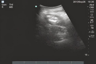
Ultrasound  abdomen first  showed  that  fluid collection around  liver and pelvis with one mass size of 3cm-4cm   at the painful area  (left lower abdomen)  like pseudokidney sign.


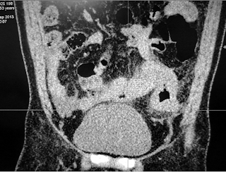
MSCT  without CE of abdomendetected  one mass with  intraluminal air and its wall was thickened more than 1cm which suggested inflammation like enteronecrosis.


This patient promptly was sent to BINH DAN HOSPITAL.and  abdomen x-ray for  check up  was done [see  photo].


Blood tests=   WBC rising 17K with  88% neutrophil.

Emergency operation as  diagnosis about peritonitis.due to perforation.
This mass is of  small intestine.which was looked like tumor or inflamation.

.
Wait  for  microscopy report.
Microscopy reports that a cancer But we have to wait for immunohistostainning to make sure that a malignant GIST or Carcinoid.

REFERENCE


CASE 211: PRESACRAL EPIDERMOID CYST, Dr PHAN THANH HẢI, Prof VÕ VĂN THÀNH, Prof NGUYỄN SÀO TRUNG, MEDIC MEDICAL CENTER, HCMC, VIETNAM

$
0
0

MALE 30 yo IN ROUTINE SCREENING CHECK-UP BY ULTRASOUND WHICH DETECTED ONE RETRO- URINARY BLADDER  MASS  WITH CYSTIC  SEPTATION.
 
MSCT  WITH  CE  SHOWED THAT MASS WITH SIZE OF 10CM,  BILOBAR, DENSITIES OF 2 LOBES  WERE  DIFFERENT. SACRUM  WAS  ERODED BY TUMOR, CONTRAST ENHANCED  PHASE  WAS VERY LOW (SEE 4  CT  IMAGES).  


OPERATION  WAS DONE FOR  REMOVING  CAPSULATED TUMOR WITH  SEBUM CONTENT LEAKING OUT [SEE  FOTO].


Pathology report is epidermoid cyst.
 
 
References  2 files pdf


CASE 212: APPENDICEAL ABSCESS, Dr NGUYỄN ĐỨC DUY LINH, Dr NGUYỄN NGỌC XUÂN GIANG, Dr PHÙ VĂN TUỐT, BÌNH AN HOSPITAL, VIETNAM

$
0
0

Man 38 yo with chief complaints: fever and lower abdominal pain for 10 days. Pain not releasing but getting worse with colicky pain and having dysenteric syndrome. Endoscopy  ruled out a  colonic tumor and noted  that sigmoid colon may pushed by an uncertain mass from outside.


Ultrasound of abdomen detected an abcess  in  minor pelvis which was thought to be an appendiceal abscess. 


Later  abdominal CT confirmed a right pelvic abscess with fecolith and a diagnosis of appendiceal  abscess was made.\
WBC= 14.76 K with 78.3% of neutrophile and CRP=87.24mg/L.


 Endoscopic surgery was performed. 
A wall-off abscess at pelvic region was detected which was due to perforated inflammed appendix at liberal tip . The pelvic abscess was removed and patient getting well.


CASE 213: BILOBED GALLBLADDER, Dr PHAN THANH HẢI, MEDIC MEDICAL CENTER, HCMC, VIETNAM

$
0
0

Woman29 yo,  recurrent pain atsubhepatic area.
Ultrasound detectedstone in gallblader with thickening wall and one cystic mass withsize of 2 cm  nearby. Sonologist reported that wasCBD cystandchroniccholecystitisdue to stone [3 images].


MSCT  also had samereport as ultrasound report (see  image CT).


Surgeonordered  MRCP. The MRI report was different which thought about  bilobar gallbladder that had 2 parts: one part had stone and  thickeningof the wall, and another part wasconnected to the neckof gall bladder which had  normalwalland normal CBD.


Endoscopic surgeryforremoving gall bladderwithstone in its fundus.
Macroscopic specimen of this gallbladder wascorrelated with 3 modalities of imaging: ultrasound, CT and MRI.


 REFERENCE


CASE 214: ADRENAL TUMOR, Dr PHAN THANH HẢI, MEDIC MEDICAL CENTER, HCMC, VIETNAM

$
0
0

Woman 50 yo, hypertension on medical treatment, well controlled, one week ago she was getting very weak and  fallingdown tobrain trauma. CTscan the head has nothing, but  blood test  showed hypokalemia  K = 1.7 mEq /ml, abnormal EKG  with  wave T= Udeformation. Ultrasoundscanof abdomendetectedone  2cm smalltumorat right adrenal area (2 pictures).


MSCTwith CE showed  this tumorinhighenhancement (HU= 8.2  rising to59.9)(see 2 CT pictures).


Blood tests fromadrenalgland hormones werenormal,aldosterone dynamictestwas=84 pg/mL,aftertest115pg/mL.
This case was operated by endoscopy for removing this tumor. See macro and microscopic specimen and report is adenoma of cortical adrenal (photo).


CASE 215: PELVIS TUMOR, Dr PHAN THANH HẢI, MEDIC MEDICAL CENTER, HCMC, VIETNAM

$
0
0
Woman 50 yo amenorrhea 3 years ago, and hypogastric area distention like being of pregnancy for 6 months.
Ultrasound at the pelvis had a masss of 20 cm in diameter, cystic septation structure which cannot separate with cervis uterus by TVS ultrasound.


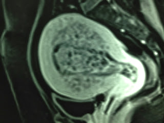
MRI of pelvis showed  that mass was cystic septation with very thick border (see MRI images).


DIFFICULTY FOR DIAGNOSING THIS CASE AS THIS MASS WAS TOO BIG, ULTRASOUND WAS LIMITED OF ANGLE OF FIELD OF VIEW.
MRI CANNOT STUDY THE MOTION OF THIS MASS, STRUCTURE WAS LOOKED LIKE OVARIAN CYSTIC TUMOR, BUT MRI  SHOWED  THE BORDER VERY THICK.
OPEN OPERATION FOR REMOVING THE UTERUS AS A SAME MASS.
SECTION OF THIS MASS WAS UTERINE FIBROMA IN NECROSIS, AND MICROSCOPY CONFIRMED.

IT IS A HUGE UTERINE FIBROMA NECROSIS LOOKED LIKE OVARIAN CYST.


CASE 216: MULTIPLE INTRAABDOMINAL NODULES, Dr PHAN THANH HẢI, MEDIC MEDICAL CENTER, HCMC, VIETNAM

$
0
0
Man 32 yo,abdominal pain, no fever. Emergency ultrasound of abdomen ruled out acute appendicitis, but there are ascites, and many small round nodules of  average of 2cm in diameter at great curvature of stomach, intra great epiploon, and at hilus of liver . All of them were  very poor echo like cyst (SEE 4 IMAGES= H.1: black nodule in hilus liver, H.2: cross section of abdomen and nodule intra great omentum, H.3:  echo poor nodule at great curvature of stomach, H4 : ascites at pelvis).


Blood test are normal. Punction of ascites fluid for analyse. PCR of tuberculosis is negative.

THIS CASE  UNDERWENT BIOPSY VIA LAPAROTOMY SHOWING  MULTIPLE WHITE SPOTS OVER PERITONEUM, LIKED CARCINOMATOSIS.


REMOVING ONE BIG MASS.AT GREAT CURVATURE OF STOMACH. CUTTING SURFACE SHOWED FLUID LIKED CASEUM.


SUGGESTION OF TUBERCULOSIS. WAIT FOR MICROSCOPY REPORT.

Microsopic  report  is  tuberculosis lymphadenitis (photo).


Discussions:


Why the result  of analysis of ascites fluid is negative  from TB PCR, ADA?
WHY
TUBERCULOSIS LYMPHNODE  are VERY  BLACK in echogeneicity?
HOW to DIFFERENTIATE it WITH LYMPHOMA ?
 

REF

.


CASE 217: THYROID TUMOR, Dr PHAN THANH HẢI, MEDIC MEDICAL CENTER, HCMC VIETNAM

$
0
0


WOMAN 41 YO, 5 YEARS AGO DETECTED SWOLLEN RIGHT SITE OF THE NECK.
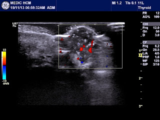
ULTRASOUND DETECTED  A RIGHT LOBE OF THYROID, SOLID,  WELL- BORDERED, WITH SIZE OF 3 - 4 CM IN DIAMETER WHILE THE LEFT LOBE WAS ATROPHY. ON  COLOR DOPPLER  IT WAS NOT A  HYPERVASCULAR NODULE.


ELASTOSCAN WAS SOFT TUMOR.


NO CERVICAL LYMPH NODE DETECTABLE
BLOOD TESTS WERE NORMAL  VALUE OF TSH , T3, AND T4.
WHAT IS YOUR SUGGESTION,  FNAC OR NOT FOR THIS CASE.

Ultrasound guided for FNAC this mass and the result was colloidal goiter.
DISCUSSION:
B-mode ultrasound of thyroid is not full examination of the thyroid, only about the thyroid anatomy .Color Doppler explaines the vasculature, and ELASTOSCAN can guide for FNAC with high accuracy.

REFERENCE


CASE 218: LEFT SUPRACLAVICULAR MASS, Dr PHAN THANH HẢI, MEDIC MEDICAL CENTER, HCMC, VIETNAM

$
0
0

MAN 55 YO, LUMBAGO FOR 3 MONTHS.  MRI  SPINAL  SHOWED SOME  DESTRUCTIVE  SPINES SUSPECTING  METASTASIS  AND  A BIG  MASS  ON  LEFT  SUPRACLAVICULAR AREA.
ON ULTRASOUND  B MODE,  IT WASA BILOBED MASSWHICH HAS DIFFERENT  ECHOSTRUCTURE . 


COLOR DOPPLER PRESENTED THIS HYPOVASCULAR MASS  LIKEDSANDWICHSIGN.


ON ELASTOSCAN  ONE  PART OF THIS MAS WAS  SOFT  AND  ANOTHER ONE  WAS  HARD MASS.


WHERE DO YOU GUIDE  FOR  FNAC (HARD MASS OR  SOFT  MASS)?
WE DID PERFORM FNAC OF THIS SOFT MASS, REMOVING EASILY THE FLUID LIKED MILK;


FLUID ANALYSIS  : GRAM STAINING= NEGATIVE BACTERIA, BK NEGATIVE, BUT CELL BLOCK WAS ADENOCARCINOMA [SEE REPORT].

CASE 219: RETROPERITONEAL TUMOR, Dr PHAN THANH HẢI, Dr NGUYỄN VĂN CHÍ, CHILDREN HOSPITAL 1 and MEDIC MEDICAL CENTER, HCMC, VIETNAM.

$
0
0
Girl 6 yo, recurrent abdominal pain. Ultrasound detected one ovoid mass at the head of pancreas with size of 3x5cm, solid and well-bordered.

Image 1: this mass and IVC.


Image 2: CDI relation with right renal hilus.


Image 3: this mass expanding artery and renal vein.


Image 4:echo structure this mass is inhomogeneous, microcalcification and
hypovascular supplying.


MSCT  with CE: clear borders and  location of  this mass.


Blood tests were normal .

What is your suggestion based on  ultrasound and CT images?


PRE-OPERATION  THERE WERE  3  SUGGESTIONS OF DIAGNOSIS, AS  RETROPERITONEAL TUMOR, ADRENAL  GLAND TUMOR AND PANCREATIC TUMOR OF  PROCESSUS  UNCINATUS.
THIS CASE  WAS OPERATED REMOVING  THIS TUMOR EASILY.  IT  HAD APPEARANCE OF HEAD OF MEDUSA  (SEE  FOTO). THE STRUCTURE  INSIDE THE TUMOR WAS IN  RED COLOR  LIKED  BONE MARROW TISSUE.


MICROSCOPIC REPORT IS NEUROBLASTOMA.


CASE 220:SMALL BOWEL G. I. S T.: Dr LÊ THANH LIÊM, Dr VÕ NGUYỄN THÀNH NHÂN, MEDIC MEDICAL CENTER, HCMC, VIETNAM

$
0
0


Male 65 yo, occult blood stool  (+) in check-up,  then underwent  colonic endoscopy to confirm colon tumor, but only colonic polyp was detected.


Ultrasound pre endoscopy disclosed  a hypoechoic mass in LLQ, semilunar shape, with size of 28x25mm which  one part of contour was regularly round and another part close by lumen gas inside a loop of small bowel . There was vessels into this mass. 


The LLQ mass was thought to be a GIST of small bowel.
CEA rising of 5.38ng/mL
MSCT confirmed  the small bowel GIST later.


Surgery was done, macroscopic result was mural tumor of small bowel


Microscopy and immunohistostaining were proved for GIST tumorof small bowel.

CASE 221:ELASTOSCANNING of FACIAL SKIN SPOT, Dr PHAN THANH HẢI, MEDIC MEDICAL CENTER, HCMC VIETNAM

$
0
0
MAN 59 YO, 3 MONTHS AGO THE FACIAL SPOT GETTING GROWTH FASTLY  AND BLEEDING. THERE WERE 2 SPOTS ON RIGHT AND ANOTHER ONE ON LEFT SITE. [SEE IMAGE 1,2].

HIGH FREQUENCY ULTRASOUND WITH ELASTOSCAN SCANNING HAD  DEEP PENETRATION UNDERSKIN  AND HARDER PATTERN WHICH SUSGGESTED BASAL CELL CARCINOMA INVASIVE. [ULTRASOUND IMAGES 1,2,3 BELONGED  LEFT SPOT, IMAGE 4 WAS RIGHT SPOT].


Biopsy the mass, microscopic report  is  BCC  (Basal cell carcinoma).


Discussion: Elastoscan map preop of  skin lesion  is  best evaluation for guiding of  biopsy and planning operation  .

REFERENCE:.Elastoscan of skin.


CASE 222: LIVER MULTIPLE SPOTS, Dr PHAN THANH HẢI, MEDIC MEDICAL CENTER, HCMC, VIETNAM

$
0
0


Man 31 yo  onset fever one month ago but not control by antibiotic. Pain in liver region.
Ultrasound  detected  some round spots  like  liver abscesses.(US  B mode, CDI, Elasto).


MRI  with  gado CE detected  also  many  round  spots,   same size of  2 cm, with central necrosis looked like metastasis.


Blood tests=   WBC rising of20k with 85% neutro, negative sero amibe, normal CEA.


Colonoscopy for detection of primary cancer disclosed, at splenic angle of colon, infiltrated indurationlesion which was suspected colon tumor .


 And biopsy with microscopic report was colon cancer.


Summary; this case is colon cancer metastases to liver.   

CASE 223: BIG IVC, Dr PHAN THANH HẢI- Dr NGUYỄN KIM THÁI, MEDIC MEDICAL CENTER, HCMC, VIETNAM

$
0
0

MAN  33 yo  PECTUS EXCAVATUM, ONSET OF  EPIGASTRIC PAIN  WITH  RADIATION  TO BACK  DORSAL SPINE.


ULTRASOUND OF ABDOMEN  DETECTED  BIG IVC  WITH  SIZE  OF =2.4-2.7cm,  ECHOGENIC  BLOOD FLOW.


SPECTRUM DOPPLER OF IVC SHOWED TRIPHASIC PATTERN WITH PV  11.2cm/s.


TEE 3D  CARDIAC  REPORT WAS NORMAL.


WHAT IS  CAUSE  OF BIG  IVC ?

MSCT of abdomen showed that IVC  is dilated in  going to heart,  diameter of 2.9-3.3 cm.


What is your suggestion ?.

CASE 224: LEFT SUPRACLAVICULAR MASS, Dr PHAN THANH HẢI, MEDIC MEDICAL CENTER, HCMC, VIETNAM

$
0
0

Man 41 yo,cough  and  pain at left arm; chest  x-rays suspected lung tuberculosis, then he had been treated as antituberculosis  for 3 months. 


Butthe pain wasgettingworse and  one massappeared at  left supraclavicular  fossa.
X-rays  again  showed this  first ribin erosion on the left site.


Ultrasoundscanning at leftsupraclavicular fossa and disclosed asolid  mass in calcification


 and fluid collection onthe rightsite.


MSCT with  CE  of the lungsdetectedthe massdestructingthe first rib. 


Biopsy was done.  

Biopsy  microscopy report  is   adenocarcinoma , it is PANCOAST TOBIAS  SYNDROME.

Ref  PANCOAT -TOBIAS SYNDROME: picture  history of Dr  PANCOAST K Henry.


CASE 201: ULTRASOUND FIRST of R. HYDRONEPHROSIS, Dr PHAN THANH HẢI, MEDIC MEDICAL CENTER, HCMC, VIETNAM

$
0
0
Woman 37 yo in pregnancy for 7 weeks, onset acute pain at rightkidney.
Ultrasound first at  FV hospital revealed  hydronephrosis of  right kidney in first degree, but cannot find out any stone. After 24 hours,  second ultrasoundat MEDIC CENTER detected one small stone  intramural urinary bladder at right ureteral orifice (see  picture 1:  hydronephrosis of right kidney , picture 2:  uterus in gestation, picture 3: intramural stone).


Urologist  requests  MRI  for make sure the right ureteral stone (2 MRI pictures).


Cystoendoscopy in emergency for releasing pain by JJ stent in ureter.
Discussion: Ultrasound firstor secondtime are better for patient by safety and cost-benefit.

CASE 202: BOWEL OBSTRUCTION, Dr PHAN THANH HẢI, MEDIC MEDICAL CENTER, HCMC, VIETNAM

$
0
0

Woman  88yo  suddenly got  epigastric pain and  vomitting. Ultrasound abdomen first  nothing abnormal detected. After 10 hours,  ultrasound in second time shows small intestine dilated and hyperperistalsis, colon no dilated (see 2 pictures of small bowel dilated) but cannot  make sure why small bowel in obstruction.


Chest X-ray  revealed air-water level at  right subdiaphagmatic, so suspected  a  subdiapragmatic abcess.

But  MSCT  detected  small intestin moving over the liver and fixed to right diaphragm and in strangulation (see CT pictures).


Summary: Ultrasound, X-ray, CT cannot make sure why small bowel in obstruction, but  surgery in emergency is done.

Emergency operation detected  small bowel fixed onto falciform ligamentum by one orifice of 2 cm diameter. It is an internal hernia due to defected falciform ligamentum. See picture of orifice of falciform ligamentum.

REFERENCE:

CASE 225: LEFT RENAL MASS, Dr PHAN THANH HẢI, MEDIC MEDICAL CENTER, HCMC, VIETNAM

$
0
0

Woman 32 yo in health care check-up:  ultrasound of abdomen detected  left renal mass in hyperechoic central  kidney with size of 2.5 cm, irregular border, and  hypovascular. Elastoscan  showedthathard mass.


MSCT with CEof thismassis in rapidenhancement andquickly washed-out.


What is yoursuggestion?

CASE 226: UTERINE MASS: Dr PHAN THANH HẢI, MEDIC MEDICAL CENTER, HCMC, VIETNAM

$
0
0

Woman45 yo,  nulliparous,vaginal bleeding.
Ultrasound  of pelvisshowed  big uterus and  thickening of endometrium.


Elastoscandetected  themassof  intrauterine cavityissoft andmultiple white spots  in myometrium.


MRI  SUSPECTED  INTRAUTERINE  POLYP. 


Biopsy result was endometrium hyperplasia. Hysterectomy was done. See macroscopic photo in correlation with ultrasound and MRI appearances.

CASE 227: Umbilical Tumor, Sister Mary Joseph Nodule, Dr PHAN THANH HẢI, MEDIC MEDICAL CENTER, HCMC, VIETNAM

$
0
0


Woman 74 yo with chief complains of anorexia, colicky pain for one month. Ultrasound detected ascites and left pleural effusion, and at  periumbilical area, there was an umbilical tumor with size of 1.5cm, solid and low vascularity.


MSCT of abdomen also doesn’t find any intra-abdomen mass, but the umbilical tumor was very high CE enhancement.


What is your suggestion  and your next step ?

Viewing all 624 articles
Browse latest View live