Quantcast
Channel: VIETNAMESE MEDIC ULTRASOUND
Viewing all 624 articles
Browse latest View live

CASE 190:LARGE TRAUMATIC PSEUDOCYST OF PANCREAS IN ADULT: Nguyen Duc Duy Linh, MD - Nguyen Ngoc Xuan Giang, MD – Dr Phan Thanh Hai, Binh An Hospital, Kien Giang, Viet Nam.

$
0
0
 A 29 year-old male patient complains:  for six months bloating of the abdomen, non deep ache, difficulty in eating and digesting food and having a mass of 150x200mm in his epigastric region. He had a trauma of epigastric region by traffic accident seven months ago and had been operated for it.
Ultrasound  examination : Cross-sectional images of the mass in epigastric region. It was a large anechoic mass with posterior acoustic enhancement, smooth contour, unilocular, no Doppler signal, size of 145x134mm, was thought to be a pseudocyst which had pressed on nearby organs (liver, stomach). It was a pancreatic pseudocyst but having a differential diagnosis of liver cyst.


MSCT examination:
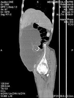
 MSCT showed a well-defined unilocular pseudocyst in the pancreatic head and body, thin wall, size of 85x138mm.


Operation:
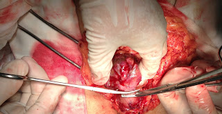
It was pancreatic pseudocyst, wallthickness of 7mm,filled yellowish fluid. Surgical drainage of the pseudocyst, which involves making a connection between the cyst and thejejunum (Roux-en-Y anastomosis).


Microscopic report:
 Pancreatic pseudocyst.


Discussion:

Pancreatic pseudocyst caused rarely by trauma and frequently happens in children. This case was a  large pancreatic pseudocyst in adult due to trauma. Ultrasound was confused with a liver cyst and priority of was MSCT higher than. In this case, surgery asked for fluid analysis but forgetting of counting amylase enzyme in withdrawn fluid, but we had microscopic report of specimen to confirm a pancreatic pseudocyst.

References

Dapo Popoola, Mary Ann Lou, and Edward H. Sims. Traumatic Pancreatic Pseudocysts .J Natl Med Assoc. 1983 May; 75(5): 515–517.

Griffith, Antonio, Wong, Lee Chu, Levine, Ho, Paunipagar. Expertddx ultrasound. Amirsys. 2010. Section 5:2-3.

Hassan A El Musharaf, Mohamed A Al Auriefi. Traumatic pancreatic pseudocyst. The Saudi Lewis G, Krige JE, Bornman PC, Terblanche J. Traumatic pancreatic pseudocysts. Br J Surg. 1993 Jan; 80(1):89-93.

Louis R Lambiase, MD, MHA; Chief Editor: Julian Katz, MD. Pancreatic Pseudocysts . http://emedicine.medscape.com.

Michael AJ Sawyer, MD; Chief Editor: Eugene C Lin, MD. Pancreatic Pseudocyst Imaging. http://emedicine.medscape.com.


CASE 191: MEDIC RADIOLOGY CASE 06:TWO CASES of CERVICAL MYELOPATHY and SPINAL SCHWANNOMA, Dr NGÔ TẤN HÙNG, MEDIC MEDICAL CENTER, HCMC, VIETNAM

$
0
0
                      
CASE 1: 47yo M patient, Vinh long province. 6 months with: Lower extremities weakness and numbness. Neck pain, muscles atrophy. No disturbance in sphincter tone as it related to his bowel or bladder function.Tender reflex (++), Babinski’s sign (++), Hoffmann’s sign (++).
MRI= Intra-and extradural and paravertebral mass of C3.
Microscopic result: Schwannoma of nerve sheath.


CASE 2 : 53yo F patient, Binh thuan province. 4 months with: Right leg weakness. No neck pain. No disturbance in sphincter tone as it related to his bowel or bladder function. Tender reflex (++), Babinski’ s sign (++), Hoffmann’ s sign (++).
MRI= Intra-and extradural mass of C6-7
Microscopic result: Schwannoma of nerve sheath.


DISCUSSIONS:
LOW EXTREMITIS WEAKNESS :  BE CAREFUL WITH CERVICAL SPINAL CORD LESIONS.


CERVICAL MYELOPATHY: A routine neurological examination is important. Complaints involving gait, equilibrium, and /or paresthesias, extremities weakness or numbness. Cervical spine pain is rarely among these complaints. [Bucy PC, Heimburger RF, Oberhill HR. Compression of the cervical spinal cord by herniated intervertebral discs. J Neurosurg.2009].


CASE 192: AVM IN THE KIDNEY. Dr Nguyen Nghiep Van , Dr Nguyen Hoai Thu, Medic Medical Center, HCMC.

$
0
0
A 51 yo female patient suffered from hematuria for a few weeks. She went to Medic Center for abdominal ultrasound. Color Doppler of the abdomen detected an AVM at the upper pole of the right kidney, d= 4.6cm in diameter, which has aliasing inside and spectral waveform of AVM (see 2 images).


MSCT with CE detected A-V shunt at the upper pole of right kidney (see 3 images).


The patient underwent DSA to make sure AVM diagnosis. The right renal artery divides into 2 branches: the upper branch feeding for lower pole of the kidney, and the lower branch, for  the A-V shunt. This is not detected on the Angio MSCT ( see 3 images ).


Noted the IVC dilated and early filling contrast, therefore we decided not to treat by coiling, because the coil may move to right atrium. And the patient transmitted to Binh Dan hospital for nephrectomy.

CASE 193: MEDIC RADIOLOGY CASE 07: ACQUIRED TRACHEOBRONCHOMALACIA, Dr LÊ HỮU LINH, MEDIC MEDICAL CENTER, HCMC, VIETNAM

$
0
0
A 31 yo female patient came to Medic Center for loss of her voice and dyspnea. For 2 years she had got suddenly deafness after giving birth. During 6 months, she coughs slightly and complaints dyspnea, getting worse when  making every effort.  There are slight whistles of her chest in 2 phases of breathing which are more clear in fast breathing.


Laryngoscopy showed normal appearance and normal motion of vocal cords, but no sound in speaking.


Bronchial endoscopy was done easily, lumen were smooth and soft, no obstacle, but getting stenosis in first part of  trachea and  bronchii which were thought to be a tracheobronchomalacia.


Chest CT confirmed  the result of bronchial endoscopy that an unknown stenosis of  1/3 upper part of trachea, stenosis of right and left main bronchii, and right intermediate bronchiole. And an old scar of right lung apex.


Because of dyspnea getting worse so she underwent a tracheostomy. After tracheostomy, chest CT proved total stenosis of trachea, and stenosis of right and left main bronchii, and  right intermediate bronchiole.


Surgery was performed to repair the trachea. And the final diagnosis is an acquired tracheobronchomalacia which causes stenosis of trachea and main bronchii. For the cause of this case is still unknown and with the deafness we may think about the immunologic reaction of a polychondritis.


CASE 194: ASCARIS in CBD and ULTRASOUND and ERCP, Dr LÊ THỐNG NHẤT, Dr TRẦN NGÂN CHÂU, Dr PHAN THANH HẢI, MEDIC MEDICAL CENTER. HCMC, VIETNAM

$
0
0
A 33 yo  female patient from Phu quoc province came to Medic Center for  painful cramp episodes 2 days before as failed in treatment of gastritis.
Ultrasound at Medic revealed an ascaris that was moving inside  common bile duct while unfortunatly CT cannot see it.


ERCP was performed to make clear diagnosis of ascaris in CBD and removing ascaris out.


The worm was still alive outside patient body being a male ascaris with hook and genital specula.


In ten years, there were 42 worms of CBD ascaris from 54 cases detected by ultrasound which were removed out by ERCP.

CASE 195: ULCERATIVE COLITIS MIMICKING COLON TUMOR, Dr PHẠM THỊ THANH XUÂN-Dr PHAN THANH HẢI, MEDIC MEDICAL CENTER, HCMC, VIETNAM

$
0
0
Male patient 26 yo suffered from colicky pain at RUQ for 2 years,  and  had a feeling of a moving mass but with only loose stool. His status is getting down.

Ultrasound detected thickening of right colon of 8 mm, and intussusception of right colon at some times. There was a walled off at right flank, so cannot ruling out a infected tuberculosis.


Colonoscopy was done but two times met intussusception, and another time in thought to be of ulcerative colitis.


Blood tests were in normal values, no clue of tuberculosis.


But MDCT detected a colon tumor with colon thickening wall of 20mm, caught CE more and loss of fatty tissue surrounding


Hemicolectomy was done by endoscopic colonoscopy, and final diagnosis is ulcerative colitis which induced intussusception of right colon.


CASE 196:PSEUDOMYXOMA PERITONEI due to Appendiceal Mucinous Adenocarcinoma, Dr. Phan Thanh Hai, Dr. Le Tu Phuc, Dr. Le Thong Nhat, MEDIC MEDICAL CENTER, HCMC, VIETNAM

$
0
0
A 75 year-old man, without history of interested diseases or surgery, came to Medic Diagnosis Center because of progressive abdominal distention for years.


Abdominal ultrasound detected an amount of large volume, echogenic ascites looked like jelly in peritoneal space. But there were some differences in morphology of ascites between right and left side of abdomen. 
In the right lower quadrant, there were two rim-calcified cysts which were adjacent to cecum. One cyst was ruptured and from this ruptured hole, many echogenic bands radiated to jelly ascites like "sunrise" in appearance. The ascites was immobile.


While in the left abdomen, the fluid was mobile with many floating echogenic nodules. Ultrasound was also detected a membrane covering small bowel loops in the left side.


MDCT showed massive ascites into peritoneum of  fat density. The ascites compressed the visceral liver surface, and the small bowel loop was pushed into the center of fluid cavity. There were two rim calcified cysts in the right lower quadrant, and one cyst had discontinuous wall. 


Blood test raised up of Beta 2 Microglobuline of 2,238 ( < 2000 Micro g/L ) and CEA of 7.83 (<5 ng/ml )

An open abdominal surgery was done at Binh Dan Hospital, removed about 5 liters of jelly-like substance. The surgeon detected a tumor of appendix adjacent to the cecum. He also reported about the membrane cover the bowel loops.


PATHOLOGY: Appendiceal Mucinous Adenocarcinoma


QUESTION: 
Why was the fluid in left abdominal side mobile but in the right one immobile?
How do we explain the membrane covering the small bower loops in the left abdominal side?

CASE 197: MIDGUT VOLVULUS, Dr LÊ THANH LIÊM, MEDIC MEDICAL CENTER, HCMC, VIETNAM

$
0
0
A 23 yo male patient came to Binh An hospital in Kien giang province with severe pain at his bossom  for  2 days, no diarrhea nor fever.  In last year he suffered some abdominal pain crisis in some times and the pain went away soon with or without medicine drugs.

In present time he cannot lie down and has to be in sitting position to reduce his pain and the pain getting prolonged.


Contrast-enhanced CT showing wrapping of the superior mesenteric vein around the SMA. 

Ultrasound detected edema of mesentery, no thickening of bowel wall and no free fluid. But the superior mesenteric artery SMA twisted itseft at  lower portion and having still flow. CT showed a case of midgut volvulus with contrast-enhanced CT showing wrapping of the superior mesenteric vein around the SMA, but with unclear cause. Plain film X-Ray revealed an intestinal obstruction. 
Surgery was done for removing twisted bowel due to an adhesive band without history of abdominal operation before. After removing the adhesive band and the twisted bowel, entire small bowel turned back in normal color.


Post-Op Diagnosis: Midgut Volvulus by adhesive band.

Xem SIÊU ÂM XOẮN RUỘT NON


CASE 198: INTRAGASTRIC POLYPOID TUMOR, Dr PHAN THANH HẢI-Dr PHẠM THỊ THANH XUÂN, MEDIC MEDICAL CENTER, HCMC, VIETNAM

$
0
0
Woman 61 yo with  epigastric distention.
Adominal ultrasound detected one  polypoid  mass being intragastric lumen. The gastric wall is rounding (4 ultrasound pictures= image 1: section of antrum, image 2 : long section over aorta,    image 3 and 4  at gastric fundus).


MSCT with 2 images, that  well show an intragastric big tumor.


Endoscopy confirmed a big polyp from gastric fundus, which has a short but large root, and rough surface, with size of 4x4 cm.


Pedunculus of gastric fundus area. Biopsy report  is adenoma.
Wait for surgery.

CASE 199: A PSEUDOANEURYM of STAB WOUND, Dr NGUYỄN NGHIỆP VĂN-Dr NGUYỄN HOÀI THU, MEDIC MEDICAL CENTER, HCMC, VIETNAM

$
0
0

A 20 yo female patient got a stab wound at her left forearm 2 months before. She presented a swellingpulsatile mass at her left forearm. Color Doppler ultrasonography shows  a pseudoaneurysm at the middle part of the left ulnar artery, d =3.2 x 3.7cm in diameter. ( see 3 images)
 

ANGIO detected pseudoaneurysm at the middle part of left ulnar artery. ( see 3 images)


Left upper extremity angiography : The half-moon images  of contrast material filling showed in the left ulnar artery.


Because the sac had small pedicle (seen on Color Doppler ), we decided to embolization the pseudoaneurysm by histoacryl glue. After embolosclerotherapy, the pseudoaneurysmal sac disappeared.


CASE 200: A LEFT BREAST MASS, Dr PHAN THANH HẢI- Dr JASMINE THANH XUÂN, MEDIC MEDICAL CENTER, HCMC, VIETNAM

$
0
0
Woman 59 yo,herself detected a mass at her leftbreast, no pain, no hot. Ultrasound findings are  a 3 cmmass under skin, noborder, hyperechoic with central necrosis, doppler no hypervascular and without axillary nodes.


On mammography, the left breast had a small nodule in hyperdensity withoud calcification and well-bordered.

What is this ?

CASE 201: ULTRASOUND FIRST of R. HYDRONEPHROSIS, Dr PHAN THANH HẢI, MEDIC MEDICAL CENTER, HCMC, VIETNAM

$
0
0
Woman 37 yo in pregnancy for 7 weeks, onset acute pain at rightkidney.
Ultrasound first at  FV hospital revealed  hydronephrosis of  right kidney in first degree, but cannot find out any stone. After 24 hours,  second ultrasoundat MEDIC CENTER detected one small stone  intramural urinary bladder at right ureteral orifice (see  picture 1:  hydronephrosis of right kidney , picture 2:  uterus in gestation, picture 3: intramural stone).


Urologist  requests  MRI  for make sure the right ureteral stone (2 MRI pictures).


Cystoendoscopy in emergency for releasing pain by JJ stent in ureter.
Discussion: Ultrasound firstor secondtime are better for patient by safety and cost-benefit.

CASE 202: BOWEL OBSTRUCTION, Dr PHAN THANH HẢI, MEDIC MEDICAL CENTER, HCMC, VIETNAM

$
0
0

Woman  88yo  suddenly got  epigastric pain and  vomitting. Ultrasound abdomen first  nothing abnormal detected. After 10 hours,  ultrasound in second time shows small intestine dilated and hyperperistalsis, colon no dilated (see 2 pictures of small bowel dilated) but cannot  make sure why small bowel in obstruction.


Chest X-ray  revealed air-water level at  right subdiaphagmatic, so suspected  a  subdiapragmatic abcess.

But  MSCT  detected  small intestin moving over the liver and fixed to right diaphragm and in strangulation (see CT pictures).


Summary: Ultrasound, X-ray, CT cannot make sure why small bowel in obstruction, but  surgery in emergency is done.

Emergency operation detected  small bowel fixed onto falciform ligamentum by one orifice of 2 cm diameter. It is an internal hernia due to defected falciform ligamentum. See picture of orifice of falciform ligamentum.

CASE 203: ULTRASOUND FIRST DIAGNOSED ECTOPIC PREGNANCY, Dr PHAN THANH HẢI. MEDIC MEDICAL CENTER, HCMC, VIETNAM

$
0
0
Ultrasound first express: Women 41 yo acute abdomen pain 7 days ago cannot lay down decubitus by pain, came to MEDIC for ultrasound in emergency with patient sitting position.


Abdomen ultrasound scan showed free fluid intra abdomen like internal bleeding, uterus was big and one round mass nearby uterus with size of 4 cm. The mass with vessel central and doppler flow was looked like ECTOPIC PREGNANCY. Scanning time lasted only 5 minutes, and diagnosing is rupture ectopic pregnancy.


Emergency ambulance sent this patient to surgery hospital.

Laparotomy was done in 30 minutes to control bleeding and remove the mass of tubal ectopic pregnancy.


Conclusion: 5 minutes of ultrasound first can save the patient life.

CASE 204: LEFT HUMERUS OSTEOLYSIS, Dr PHAN THANH HẢI, MEDIC MEDICAL CENTER, HCMC, VIETNAM

$
0
0

Man 42 yo  after playing golf, got pain at  left upper  humerus.
Plain xray   showed   the humerus bone had been in  osteolysis.


Ultrasound  made sure that  cortical bone rupture of upper portion the left   humerus, without changing  periosteal  space, and arrounding muscle were intact.


CTscan with CE  reported  a suggestion  of BONE CYST ANEURYSM. Do you agree with this  suggestion or  not?


What is your suggestion?

CASE 205: MEDIC RADIOLOGY CASE 9= Noncompacted Cardiomyopathy on MSCT 640, NGUYEN TUAN VU, NGUYEN THI KIM SANG, DUONG PHI SON, PHAN THANH HAI, MEDIC MEDICAL CENTER, HCMC , VIETNAM

$
0
0

HISTORY
Female patient , 33 yo, presented by severe heart failure for  1 year, previous diagnosis : dilated cardiomyopathy . Decreased S1. audible S3,  2/6 apical systolic murmur.  She was sent to MEDIC for cardiac MSCT to rule out Coronary artery disease.
EKG
Short PR, delta waves, QS in V2-V6, D1 aVL 


ECHOCARDIO+ TDI and 3 D Echocardiography
Decreased LV systolic function , LV diastolic dysfunction , LV diastolic dysfunction
Prominent trabeculae, spongiformed LV , Diagnostic criteria NC/C leyer > 2
Noncompacted cardiomyopathy Echocardiography: Apical 4 C view and Parasternal short axis view
Prominent trabeculations and spongiformed myocardium of LV 


MS CT 640               
Intertrabecular Recesses, Multiple Trabeculae , Predominant location at Apex, mid lateral, mid inferior . NC layer/ C layer > 2,2, Involving >2 segments, Sens. 100%, Spec. 95% (Tomography, volume 6, Issue 5, Sept.-Oct. 2012, pp 346-354)
MSCT 640: 3D Imaging =Trabeculated and spongiformed LV 


 Video clip from apex view


Summary
—  Reported by Engberding and Benber in 1984 :Mutation in LDB3, genetic cardiomyopathy
—  Myocardial sinusoids
—  Severe heart failure, arrhythmias, thrombus formation, sudden death
—  Diagnosis by  Echocardiography, MRI, MSCT
—  Medical treatment ( ACEI, Betabloker, Aspirin, Anticoagulant ), ICD, heart transplant.
—  Long term prognosis is unknown
—  Value of cardiac MSCT in patient with heart failure.


CASE 206: EPIGASTRIC MASS POST- PROSTATECTOMY for 5 YEARS, Dr PHAN THANH HẢI. MEDIC MEDICAL CENTER, HCMC, VIETNAM

$
0
0
Ultrasound check up  a 62 yo man , who underwent  prostatectomy for 5 years, detected one hypoechoic epigastric mass, size of 5cm in relation with great curvature of stomach. Color Doppler showed vascular supply from gastric artery.


Gastro-endoscopy detected no lesion inside stomach.

MSCT with CE showed this tumor was from gastric wall, and pediculated.


Blood test were normal all cancer markers: PSA, CEA , CA 19-9, CA 72-4.

Operation  laparotomy..showed this tumor is from  the  great curvature of stomach   with  long pedicule. Resection tumor see  macroscopy, wait for  microscopic report.


CASE 207: GASTRIC DUPLICATION CYST, Dr PHAN THANH HẢI, Dr NGUYỄN THIỆN HÙNG, Dr TRẦN NGÂN CHÂU, MEDIC MEDICAL CENTER, HCMC, VIETNAM

$
0
0

A 26 year-old male patient  from Kien Giang province with chief complain: nausea and mild epigastric discomfort for 3 years, no vomiting, no fever. Family and his past medical history : nothing abnormal detectable. Physical examination: mild epigastric tenderness,  no mass in the epigastric area.


Undergoing of gastroendoscopy he was revealed a submucosa mass in the antrum which was confirmed  by CT; it was  a fluid-attenuation cystic mass in close  with the stomach wall but report of CT cannot rule out a heterotropic pancreas.


Ultrasound detected an 27x17mm intragastric cyst  which adhered the greater  curvature. The cyst wall had 2 layers: echogenic inner mucosal lining and hypoechoic outer rim which was contiguous with the muscular layer of the stomach. So we met a muscular rim sign of a  non-communicating GDC (gastric duplication cyst) in adult.


Operation at BD Hospital confirmed the non communicating GDC.


Wait for microscopic result.

CASE 208: RETROPERITONEAL EXTRAUTERINE PREGNANCY, Dr PHAN THANH HẢI, MEDIC MEDICAL CENTER, HCMC, VIETNAM.

$
0
0
WOMAN  26yo UNDERWENT in vitro fertilization–embryo transfer (IVF–ET) WAS WAITING FOR THE RESULT.

BETA HCG  WAS RISING UP FROM 9K  TO 17K  BUT ULTRASOUND  CANNOT FIND OUT THE   INTRAUTERINE NIDATION.

AT THE RIGHT  OVARY SITE,  ULTRASOUND DETECTED ONE  MASS OF 2 cm WITHOUT BLOOD SUPPLY WHICH WAS  BIG AND  CYSTIC HEMORRHAGIC, NEAR RIGHT ILIAC VEIN.
.


FIRST LAPAROSCOPY DETECTED NOTHING.
ONE WEEK LATER   MRI WAS PERFORMED ALSO DETECTED  THIS MASS WHICH WAS  GROWING IN RETROPERITONEAL SPACE, NEAR  RIGHT ILIAC VEIN. Beta HCG AT THAT TIME WAS  UP TO  39K.


OPEN  SURGERY  REMOVED THIS  MASS, SUSPECTED EXTRAUTERINE PREGNANCY 
WAIT  FOR   PATHOLOGY REPORT  AND  FOLLOW UP THIS CASE  AS  EXTRAUTERINE PREGNANCY IN RETROPERITONEUM.

CASE 209: A LUNG MASS, Dr PHAN THANH HẢI, MEDIC MEDICAL CENTER, HCMC, VIETNAM

$
0
0
Man 59yo in check-up lung x-ray detected a mass at left lung, asymptomatic, no history tuberculosis, non smocking patient.


Ultrasound of the lung showed that was a cystic avascular with well bordered mass, size of 4 cm.


MSCT CE said that cystic mass bilobar looked like a bronchogenic cyst.


Do you compare 3 facilities for the diagnosis of this case ?.
Viewing all 624 articles
Browse latest View live