Quantcast
Channel: VIETNAMESE MEDIC ULTRASOUND
Viewing all 624 articles
Browse latest View live

CASE 170: A BREAST TUMOR, Dr PHAN THANH HẢI, MEDIC, MEDICAL CENTER, HCMC, VIETNAM

$
0
0
Woman 77 yo, by herself detected one lump at herleft breast.
Ultrasound  examination: this mass was at  section  of 10 hr  of left breast, size  arround 2 cm ( B mode US picture). 

It was  hypoechoic and  irregular  border, with very strong shadowing (image 2 and 3), and on CDI, hypervascular and  very high PI.


On PDI again, this tumor was hypervascular; axillary scan no detected nodes.


Ultrasound first  suggests breast cancer, next step ismammography or  MRI.
 
THIS PATIENT  REFUSED  TO DO MAMMOGRAPHY AND MRI BECAUSE  THE FIRST TECHNIQUE  WAS PAINFUL  AND THE SECOND ONE MADE  CLAUSTROPHOBIA FOR  HER LONG TIME AGO.
MSCT  IS CHOSEN FOR STAGING  THIS CASE. (SEE  3 CT SLICES )


MSCT non CE  showed that tumor  was small size of  1.8 cm, spiculate hypercalcification and detected no  lymphatic nodes of axillary or retrosternum, it was staging I.
Biopsy was done and report was breast cancer type NOS.


CASE 171: THICKENING OF ANTRUM WALL, Dr PHAN THANH HẢI, MEDIC MEDICAL CENTER, HCMC, VIETNAM

$
0
0

Woman 39yo, anorexia, vomitting and loss weight rapidly.

Ultrasound abdomen first, see the dilated stomach too big, and the antrum thickening of the wall like the uterus cervix (pseudocervix sign). At the pelvis,  uterus was covered around by ascites (see 3 ultrasound pictures).

MDCT ABDOMEN WAS DONE, some FRONTAL, AND SAGITAL SECTIONS SHOWED THE ANTRUM THISKENING OF THE WALL.

GASTRO-ENDOSCOPY SAW THE ANTRUM STENOSIS.


BIOPSY WAS PERFORMED. WAIT FOR MICROSCOPY REPORT.
ALL OF THE DIAGNOSTIC PROCEDURES SPENT FOR 2 HOURS.

Biopsy report  was  gastric  cancer.

CASE 172: HEPATIC ECTOPIC PREGNANCY, Dr PHAN THANH HẢI, MEDIC MEDICAL CENTER, HCMC, VIETNAM

$
0
0

Woman 24 yo, amenorrhea for 2 weeks, was suspected  in pregnancy, but ultrasound at pelvis showed uterus without gestational sac or mass beside uterus.


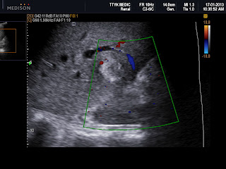
Ultrasound scan at liver detected one hyperechoic focal, hypovascular, round shape, size of 1.86 cm with fluid in central mass.


Blood test beta HCG is of 34k unit. Do you thing it is an ectopic pregnancy in liver and how to make sure the diagnosis for this case?.


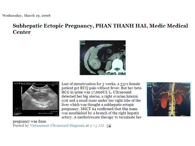
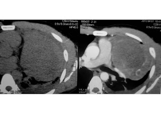
MDCT with CE  was done  for  detection  the intrahepatic focal which was  near  the gall-bladder, size of  2cm, hypodense  cystic central and  blood supply by  hepatic artery (see 3 CT images).


ULTRASOUND AND MSCT LIVER SUGGESTED PRIMARY LIVER PREGNANCY WITH high value of beta HCG 32 k unit/ml. Methotrexate is drug of choice for treatment, after 2 weeks of injection of methotrexate the blood test beta HCG will be dropped to normal, the liver focal will get smaller as a cyst. This is a case of PLP (PRIMARY LIVER PREGNANCY) succesfully treated with METHOTREXATE. NO NEED of OPERATION. IT IS RESULT OF EARLY DIAGNOSTIC of PLP.

REFERENCE: Case in MEDIC of DATE 2008: Subhepatic Ectopic Pregnancy 

CASE 173: THORACIC OUTLET SYNDROME, Dr NGUYỄN PHƯỚC BẢO QUÂN, MEDIC CENTER in HUẾ

$
0
0

Female  33 years old complains pain in right arm when she has her arm in abduction and elevated position.
Ultrasound findings:


Fig 1: Right side of the image indicates normal dimension on transversal section of the R subclavian artery (arrow) before the test by which the patient elevates her arm in external rotation; left side of the image indicates small dimension on transversal section of the R subclavian artery during the test due to compression between the anterior scalene muscle anteriorly (white arrow head) and exostosis of the first rib posteriorly (black arrow).

Figure 2:The spectrum waveform of the radial artery before and during the test. 


Fig 3: Longitudinal section of the R subclavian artery indicates the stenosed segment with high flow velocity displayed by aliasing phenomenon and  post-stenotic dilatation segment as well. Note that focal thickening of the wall of the R subclavian artery at stenotic region (white arrow).
Fig 4: CT Angio images of the R subclavian artery demonstrate the stenosed segment due to exostosis of the first rib (red arrow) and poststenotic dilatation segment.
Diagnosis: Thoracic outlet syndrome in the first space.
Discussion: Thoracic outlet syndrome (TOS) is the name of a variety of conditions attributed to compression of the neurovascular structures as they traverse the thoracic outlet. (TOS) can occur at 3 spaces: 1/ The first space is the interscalene triangle. It is bordered by the anterior scalene muscle, the middle scalene muscle, and the upper border of the first rib. The interscalene triangle is the most common site for neural compression, vascular compression. 2/ The second space is the costoclavicular triangle, which is bordered by the clavicle, first rib, and scapula and contains the  subclavian artery and vein and the brachial nerves; 3/ The third and final space is beneath the coracoid process just deep to the pectoralis minor tendon; it is referred to as the subcoracoid space.
Reference: 
 1.Daryl A Rosenbaum, MD; Chief Editor: Sherwin SW Ho, MD. Thoracic Outlet Syndrome . http://emedicine.medscape.com.
2/ Paul B. Kreienberg, Dhiraj M.Shah et al. Thoracic outlet syndrome. Vascular diagnosis. Elsevier Saunders. 2005. P.512-522


CASE 174: HCC or NOT ? Dr PHAN THANH HẢI, MEDIC MEDICAL CENTER, HCMC, VIETNAM

$
0
0

Male patient 56 yo strickly followed up by HCV, 2 weeks ago fever, chill, pain at liver region. Ultrasound of liver detected one mass at right lobe, size of 10cm with hypoechoic mixed structure inside. Doppler showed hypervascular. 2 fellows in sonology said that to be HCC.

DO YOU THING IT IS HCC WITH 5 ULTRASOUND IMAGES?.

Blood test report:


MDCT of liver without CE and with CE were in suggestion of liver tumor by radiologist report.


Do you thing ultrasound and CT can make diagnosis for this case, or clinical and blood test are the main reasons for diagnosis?.

What could you do next for this patient?.


This patient had been admitted in infectious tropical hospital. Blood test negative for amebiasis, and fasciola hepatica; antibiotic was in perfusion. After 2 days, patient was not in fever. Wait for result of blood culture and ultrasound reviewing the liver mass.

CASE 175: A CARCINOMA of THYMUS, Dr PHAN THANH HẢI, MEDIC MEDICAL CENTER, HCMC, VIETNAM

$
0
0
Man 27 yo, one month ago, got cough and dyspnea at night. Chest X-ray showed one mass of 10cm near the apex of the heart.


Chest ultrasound, in sitting position, revealed pleural effusion, like bloody and this mass on chest X-ray was solid and covered the heart.


MDCT  of the lungs  with CE,  this mass belongs to anterior mediastinum and in extention around the pericardium and pleural effusion. Radiologist suggested a teratoma.


Do you agree with the CT diagnosis ?


Thoracotomy  the  case  removed  1.7  liter of  yellowish  fluid, and  tumor looked like  brain tissue.


Microscopic result  is  suggestive of  undiffentiated  thymocarcinoma. 


REFERENCE:


CASE 176: PARATHYROID TUMOR, Dr HỒ CHÍ TRUNG, MEDIC MEDICAL CENTER, HCMC VIETNAM

$
0
0

Female patient 49 yo from Phu yen province. Slight edema, 2 leg weakness for 2 years, using wheelchair to many HCM hospitals.
She went to Medic Center in hope to make clear her illness. 

Chest X-Ray: deformation of chest bones and osteoporosis. 
Column vertebra : biconcave of vertebral bodies due to osteoporosis.

Ultrasound at Medic revealed a hypoechoic mass in the lower pole of left lobe of thyroid, and hypervascularization which was thought to be a parathyroid tumor.


PTH highly raising up of 1,048 picog/ml [16-65 picog/ml] and bone scan proved osteoporosis.
In 2 hours post op, PTH dropped down of 47.21picog/ml.

 And microscopic result= Parathyroid Adenoma.
So it is an severe osteoporosis case due to parathyroid adenoma that is unveiled successfully at Medic Center in some days while it lasted for the female patient in 2 years in her wheelchair.

CASE 177: MEDIC RADIOLOGY CASE 01= Massive Pulmonary Embolism, Dr NGUYỄN VĂN CÔNG, MEDIC MEDICAL CENTER, HCMC, VIETNAM

$
0
0
Male patient 47 yo, admitting to the clinic because of chest pain.
Chest Xray shows no significant finding  beside slightly bulging of L pulmonary artery.


ChestCT scan with contrast at Medic Center shows R pulmonary 90% occluded with thrombus and L upper lobe artery also occluded 30%.


The patient is transferred to Singapore and conservative treated because the clot is too old and well organized.

CASE 178: CHORDOMA, Dr LÝ VĂN PHÁI, MEDIC MEDICAL CENTER, HCMC, VIETNAM

$
0
0

56-year-old female patient from Tra Vinh province. She's undergoing  low back pain  more than 1 year, recently appeared constipation. Spinal x-ray shows destruction of the sacrococcygeal bone. 
 

Ultrasound shows lesion that is a  heterogeneous hypoechoic solid tumor, size 72 x42 x 62 mm, inner tumor to be calcified.


MRI shows tumor from sacrococcygeal bone, dimension = 5 x 6.5 x 7cm, high density on T2W1 and low on T1W1.


Patient underwent surgery at Cho Ray hospital. Histopathologic report is  chordoma.


CASE 179: CHRONIC DIARRHEA and CAPSULE ENDOSCOPY, Dr PHẠM CHÍ TOÀN, MEDIC MEDICAL CENTER, HCMC, VIETNAM

$
0
0

Clinical history=
Female, 91 years old, Nha be district. For 3 months: watery diarrhea without blood (over 10 times/day), no abdominal pain. Treated by many doctors and medical centers, but diarrhea did not stop.
Tropical disease hospital admission: failed treatment after a month and transmitted to Binh dan hospital.

v  Lab test: normal, Abdominal US,  CT scan: normal, UGE : normal, Rectosigmoid endoscopy: normal. Stool test: negative, suggestion : capsule endoscopy.

v  Capsule Endoscopy= Many worms with the appearance of Ankylostoma in jejunum and ileum and inflammed mucosa of small bowel.

Diagnosis=Enteritis due to parasite (maybe Ankylostoma duodenale).  Internet : some case reports like this.

Treatment=
Suggestion : stop using all kinds of drugs including antibiotics, start with Zentel 2 tablets a day/ 3 days. Diarrhea improves in getting down : 10 times and 6 timesand4 timesand 2 times. Discharge. Suggestion: repeated Zentel after a week, now normal stool.

Conclusion=

Few case reports, besides anemia, acute or chronic diarrhea sometimes happens. Easy and simple treatment.

But the worm name should be proved by ELISA test for Ankylostoma.

CASE 180: AV FISTULA POST OP MIDDLE EAR SURGERY for 10 YEARS, Dr LÊ THANH LIÊM, MEDIC MEDICAL CENTER, HCMC, VIETNAM

$
0
0
 Male patient 30 yo underwent a middle ear surgery for  10 years. After  surgery about  2 years, a posterior ear mass appeared and getting its size bigger .

 There are thrills in the mass, and it is collapsed with palpation.
 Ultrasound detects tortuous dilatation of subcutaneous vasculature at temporoparietal area on right side and around right auricular area, Doppler shows continuous  flow pattern which is thought to be an AV fistula post op.


  MDCT confirms an AV fistula on 1/2 right head which is supplied by superficial temporal artery and drains away by facial and right jugular veins.


CASE 181: APPENDICULAR MUCINEOUS CARCINOMA and INTUSSUSCEPTION, Dr PHAN THANH HẢI - Dr VÕ NGUYỄN THÀNH NHÂN , MEDIC MEDICAL CENTER, HCMC, VIETNAM

$
0
0
Woman 73 yo has  pain at  right pelvis for 6 days, crisis, hyperperistalsis. Ultrasound  shows  one cystic mass  nearby  urinary bladder, with size of 4 cm, round  border with the neck connecting to lumen of  intestine.
See 3  ultrasound pictures with 3.5 MHz curve probe, and with  linear probes 12 MHz; this cystic mass has multilayer wall, calcification of the border, the fluid is cloudy. This cystic mass  is  moving  with  hyperperistalsis due to intussusception (3 images).


On MDCT this  mass is cystic,  intra  intestine lumen,  portion of  terminal ilium.


This case has emergent onset. Ultrasound first says that intussusception, but don't know what  the cyst is; MDCT  suggests an appendicular mucocele. Do you agree with the idea ?

Operation is laparotomy which detects  right colon very mobile with one mass at appendix site.


Procedure is right hemicolectomy. In macroscopic dissection, specimen is an appendicular mucocele.  Wait for confirmation of microscopy.

The  clinical  case  of  intussusceptions  in  old  woman  has  the cause. Ultrasound   detects  cystic  mass  close to urinary bladder  which  is  not common cause  for  ileo-colic  intussusception.
 MCDT  reveals   cyctic mass  intra intestinal  lumen with  mural calcification is  the key point  for   diagnosing  this case.

Microscopic report  is mucinous carcinoma of appendix.


CASE 182: Malignant Melanoma of Choroid, Dr LÊ THANH LIÊM, MEDIC MEDICAL CENTER, HCMC, VIETNAM

$
0
0
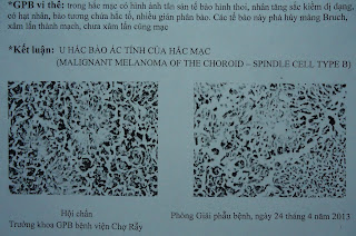
Man 53 yo,  Binh duong province, loss of right vision for 3 months,  ultrasound and OCT of  Eye Hospital think about melanoma of ciliary body.


At  Medic Center, ultrasound detects a right intraocular tumor, size of 12x10mm, at anterior and internal location, echo rich, round border, no calcification, vegetation and nor posterior shadowing.  It is a hypovascular tumor.  There is a right retinal detachment.


 On elastographic ultrasound with eSie Touch and ARFI techniques ( Acuson S2000, Siemens) velocity of VTI is hard in moderate grade, homogenous, in grey color meanwhile tumor is hard and homogenous in eSie Touch ( black and blue).


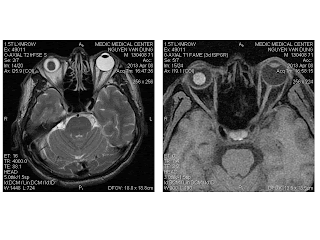
MRI reveals an intraocular tumor on right eye, which is from ciliary body, size of 12x11mm, low signal on T2W1, and high signal on T1W1 and T1GRE,  lightly catched Gado, and no extraocular invasion. Follow-up  a Choroidal-Ciliary Melanoma.

Surgery was done on 15-4-2013,  microscopic result :malignant melanoma of the choroid.


CASE 183: LEFT ARM MASS, Dr PHAN THANH HẢI - Dr LÂM CẨM TÚ, MEDIC MEDICAL CENTER, HCMC, VIETNAM

$
0
0
Man 27 yo,  3 months  ago  detected  mass  at  left  arm  ( foto)  no pain  no trauma  history and slowly  growth.

Ultrasound first  seeing  this mass  is  hypoechoic,  intramuscular, well  bordered  with  very  high  pattern color Doppler.

MSCT  with CE  of  this  mass  filling rapidly contrast  agent  that  looked  like  an A-V  MALFORMATION.

OPERATION REMOVES  EN BLOC TUMOR, WELL BORDERED WITH  OPENED SURFACE   IS CONVEX, LOOKED LIKE BENIGN TUMOR.


MICROSCOPIC WITH IMMUNOHISTOLOGIC STAINING REPORT: Hemangioendothelioma, it is  very rare soft tissue tumor.


CASE 184:MEDIC RADIOLOGY CASE 02: MEDIASTINAL LIPOMATOSIS, Dr NGUYỄN VĂN CÔNG - Dr HỒ CHÍ TRUNG , MEDIC MEDICAL CENTER, HCMC, VIETNAM

$
0
0

Male patient 54 yo takes CXR for heath check at MedicCenter.
Surprisingly we found a very enlarged heart shadow which oblitrated ½ lower chest on both PA and lateral chest XR.


On cardiac ultrasonography, the heart is within normal limits, but there are abnormal structures surrounding the heart with hyperechogeneicity.


We decided to do a thoracic CT scan to clarify the problem.


The result of CT scan shows a very large structure of  fat density occupied all lower half of the chest and confirmed by surgery and pathology: about 2 kg of fat was removed from lower mediastinum due to mediastinal lipomatosis.


CASE 185: MEDIC RADIOLOGY CASE 03: RETROCARDIAC MASS, Dr NGUYỄN VĂN CÔNG-Dr HỒ CHÍ TRUNG, MEDIC MEDICAL CENTER, HCMC, VIETNAM

$
0
0
61 yo female patient went to MedicCenter for a health check-up be cause her sister recently died from lung cancer.


PA and lateral chest XR revealed of a suspicious big mass  of 11x 5cm on L lung base : was that a tumor?
  
An ultrasound examination was performed to know that mass is solid or cystic nature. To our surprise, a typical structure of a kidney is detected by echography in ectopic situation but difficult to certified it is above or under the L diaphragm.


The problem was easily resolved by  CT scan with contrast showed nicely the L kidney well vascularized and preserved function herniated through Bochdalek foramen.


So it was an ectopic thoracic kidney and diaphagmatic hernia.                   

CASE 186: LYNCH SYNDROME OR NOT ? Dr JASMINE THANH XUÂN, Dr HỒ CHÍ TRUNG, Dr PHAN THANH HẢI, MEDIC MEDICAL CENTER, HCMC, VIETNAM

$
0
0

Female patient 28yo underwent R hemicolectomy as adenocarcinoma in November 2011 at BD hospital. On 4 April 2013 she came back to BD Hospital to be removed  both 2 ovarian tumors (Krukenberg) for metastase from colon tumor. And after surgery of ovarian tumors for 3 weeks, she detects herself her right breast swollen, hard in palpation but without pain so she returned BD hospital again.

BD hospital ultrasound showed secondary right breast tumors and she was sent to MEDIC to take a mammography.

At Medic, X-Rays detected a breast mass, with high density on RUQ of right breast and were thought to be a BIRADS 4 tumor,


and Medic ultrasound again confirmed 3 hypoechoic nodules of 13-22-8mm at 10h, 11h, 1h, irregular borders, with microcalcifications (BIRADS 4)


and right metastatic axillary nodes,


and some R and L cervical nodes.


MSCT 640 proved a right breast tumor with ROI=25HU and axillary nodes and R pectoralis muscles nodes.


FNAC of right breast tumor was done and microscopic result was adenocarcinoma.

In conclusion, a 28yo female patient which were removed R colon tumor and ovarian tumors due to metastase now has a R breast tumor  BIRADS 4 and R axillary nodes and R and L cervical lymph nodes.

Do you think a case of Lynch syndrome? Is it sure that secondary breast tumors? What to do in the next step ?


Surgery was performed on 24/5. So it was a primary breast tumor, both macroscopic and microscopic findings and not a metastatic tumor of the breast from colon tumor and that may belong to Lynch syndrome. Waiting for genetic test to confirm  the final diagnosis of Lynch syndrome.

CASE 187: MEDIC RADIOLOGY CASE 04: 170 CORONARY ARTERY CASES SCANNING by MSCT 640, Dr DƯƠNG PHI SƠN-Dr PHAN THANH HẢI, MEDIC MEDICAL CENTER, HCMC, VIETNAM.

$
0
0
      170 cases (79 male / 91 female) in our 2 weeks of setting up the new machine.
      Angina  pectoris and risks of coronary artery disease.
      Contrast agent: Ultravist, dose <1ml/kg
      Radiation dosage <1-4 mSv (DLP x K-factor =DLP x 0.014)
      Appropriate heart rate  < 70 p/min, if existing tachycardia can use Beta Blockers, Diltiazem... to slow down heart rate 
      Appropriate blood pressure under 140/90 mmHg
      Patients hold their breath during shooting.


CONCLUSIONS:
     Image quality greatly improved (increased levels of accuracy).
     Radiation dose reducing 50-90% and extremely low amount of contrast agent (competitive and beneficial to the patient).
     Cardiac arrhythmia still could be scanning (more advantage than the older generation machines).
     Shooting and find the cause of the chest pain emergency.

CASE 188: MEDIC RADIOLOGY CASE 5: THYMOMA and THYMOCARCINOMA, Dr Nguyễn văn Công-Dr Hồ Chí Trung, Medic Medical Center, HCMC, Vietnam.

$
0
0


Mr A. and Mr B. are two 64 yo male patients, have a heath check at Medic Center some days apart.


On the PA and lateral CXR a round shadow about 3-6 cm of diameter seen on anterior mediastinum R side.


On thoracic US examination, 2 encapsulated, with septations mixed structrure of solid and liquid contents on R lower anterior mediastinum of 3-5 cm diameter look alike on both patients.
          

CT scan of both chests show almost the same findings as a mixed components with solid and fluid, septated, encapsulated, contrast enhanced on R lower anterior mediastinum.

Imaging diagnosis: Right Anteroinferior Mediastinal Tumor can be thymic origin.
Both men go for surgery to remove the anterior mediastinum tumor.

 But the resullts from pathology are different:

The pathology report of Mr A.: THYMOMA TYPE A  WITH CYSTIC CHANGE. 
The pathology report of Mr B.:Undifferentiated Adenocarcinoma, invading lung tissue, EGFR (+)


Outcome of the patients:

Mr A. after the surgery is still living and working till now.
Mr B. still alive some months after radiation and chemotherapy.

A coincidence : 2 cases of incidentaloma with different outcomes. From this lesson we can learn that:

1/ Imaging diagnosis is only the shadow of the truth.
2/ Pathology give us one part of the truth.
3/ Clinical findings, imaging and pathology can give us almost the truth.
4/ But the truth itself  is the outcomes of the patients .


CASE 189: NUTCRACKER‘S SYNDROME, Dr PHAN THANH HAI, Dr NGUYEN DUC TRI, MEDIC MEDICAL CENTER, HCMC, VIETNAM

$
0
0

Male boy 12 yo had been in operation of cystostomy for removing blood clot 3 months before, but unkwon cause , and now hematuria is recurring.

Ultrasound of abdomen detected left renal vein dilated, big and big in comparison to one on right site, color Doppler shows very low flow in left dilated renal vein. (see 3 pictures).


 2 kidneys are normal.

MSCT CE UROLOGY detected left renal vein dilated and pressed between aorta and SMA,  and early filling contrast,  suspected one A-V SHUNT at upper pole of left kidney.(See 3 images of CT with CE).


Planning to do DSA for make sure diagnosis of hematuria and site of bleeding.
Viewing all 624 articles
Browse latest View live