Quantcast
Channel: VIETNAMESE MEDIC ULTRASOUND
Viewing all 624 articles
Browse latest View live

CASE 479: TOOTHPICK PERFORATED BILIARY DUODENUM, Dr PHAN THANH HẢI-Dr LÊ THỊ THANH THẢO, MEDIC MEDICAL CENTER, HCMC, VIETNAM.

$
0
0


Man 74 yo with epigastric pain, being treated like gastric ulcer.
Ultrasound of abdomen  detected  one hyperechoic foreign body # 3.5 cm which penetrated  gastro-duodenum wall  to gallbladder. The  gallblader wall is very thick # 1 cm.


MSCT of  abdomen made diagnosis  that was a toothpick  penetrated from gastric wall to  gallbladder.


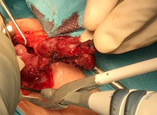
Operation removed this toothpick and cholecystectomy.


Conclusion:  Toothpick perforated gastric wall to gallbladder, an emergency case must be known.


CASE 480: BILATERAL MAMMARY-OVARIAN LYMPHOMA, Dr JASMINE XUÂN, Dr TRẦN NGÂN CHÂU, Dr PHAN THANH HẢI, MEDIC MEDICAL CENTER, HCMC, VIETNAM

$
0
0


Woman 33yo with pain in lower maxillary  for one month and tension in both of 2 breats ( see photo) without pregnancy.  


US scanning of abdomen= big cervix  of uterus,  ascites and 2 ovarian solide tumors,  size 5-7 cm (US 1, US 2). 


US of mammary scanning  showed  small  hypoechoic  nodules  infiltrating in 2 breasts without axillary node ( US 3-US 4).


US 5-US 6-US 7 =  ABVS scanning   detected  multiple nodules infiltrating in 2 breasts.


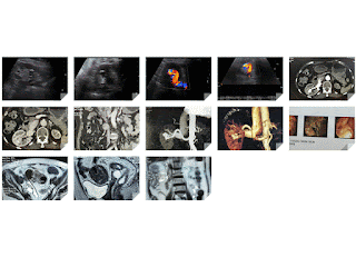
  • MRI  full body with gado detected  bone marrow changing, 2 breats  hypercaptured contrast  ( MRI 1),  ascites and kidney infiltration ( MRI 2).


MRI 3 : pelvis  with 2 ovarian tumors and big uterine cervix (MRI  4).


Blood tests=   lower platelets,  EGFR  lower  46, beta2 microglobuline raised  3816,  ferritine raised  911, LDH-l  raised  1360.
Basis clinical, imaging and blood tests  suspected  lymphoma  diffuse type.
Biopsy of 2 breasts  reported  microscopic with IHC, beta cell lymphoma.


Summary =  Lymphoma stage 4  infiltration in 2 breasts and 2 ovaries  for this case.  

CASE 481: ZENKER’S DIVERTICULUM, Dr PHAN THANH HẢI, Dr PHẠM THỊ THANH XUÂN, MEDIC MEDICAL CENTER, HCMC, VIETNAM.

$
0
0


Man  38yo with shore throat, suspected GERD but  esophago-gastric endoscopy is gastritis.


Ultrasound of the neck at left lobe thyroid detected a cyst # 1.5 cm in diameter  (US 1, US 2 ) and compression maneuver over this cyst made it smaller.


US 2:  longitudinal scanning of left lobe this cyst had air inside.
US 3:  CDI with Doppler artifact reverberation (US 4).


Sonologist report is esophageal diverticulum.
X-Rays of swallow barium showed normal esophagus.


MSCT at cervical area with iodine contrast swallow showed air in this cyst  and contrast filling inside.


Radiologist report is Zenker‘s diverticulum of esophagus.
http://www.journalmc.org/index.php/JMC/article/view/784/392

REFERENCE: Case report  of Journal of Medical Cases.


CASE 482 : SPLENOSIS, Dr PHAN THANH HẢI- Dr TRẦN CÔNG DUY LONG, MEDIC MEDICAL CENTER, HCMC, VIETNAM.

$
0
0


Man 38yo with  abdominal pain  like gastritis. Clinical detected arterial hypertension.
In past history he had been in an urgent operation of rupture of spleen by trauma for 10 years ( photo).


Ultrasound  detected  one  mass at border of right liver near upper pole of right kidney  and  sonologist suspected an adrenal gland tumor  ( US 1, US 2 CDI , US 3  view with linear probe).


MSCT of  abdomen=   CT 1:crossed section of  tumor  at border of liver, 
CT 2 with CE  is  low enhanced  tissue.


After being treated in stable blood pressure, a laparotomy removed  big tumor at liver border in retroperitoneum  and  some intra abdomen small nodules.


Microscopic report is normal tissue of spleen.


It is  splenosis  due to rupture of the spleen 10 years before.   

CASE 483: UTERINE GIANT MYOMA, Dr PHAN THANH HẢI, Dr DƯƠNG NGỌC THÀNH, MEDIC MEDICAL CENTER, HCMC, VIETNAM.

$
0
0


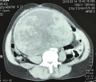
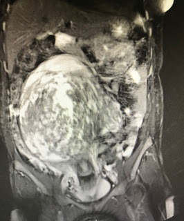
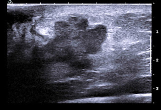
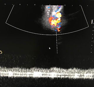
Woman 47yo  still has mense with the big growth of abdomen (photo).


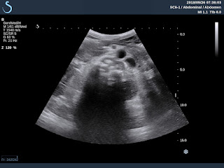
US scanning  detected  a solid tumor cover over abdominal cavity ( US 1, US 2  US 3  longitudinal scanning over aorta),  US 4 : elastoscanning of tumor structure is hard and  inhomogeneous.


CT scan with CE of this mass is enhanced CE slowly: CT1, CT 2  sagittal view of  the tumor and uterus, CT 3: tumor near right kidney.


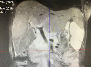
MRI  with gado= MRI 1:  crossed section, necrotic cystic formation of tumor structure. MRI 2 : longitudinal scan, MRI 3:  frontal view.


Preoperative diagnosis is big uterine myoma.
Macroscopic  photo of this tumor.


Hysterectomy was done.

CASE 484: SPLEEN TUMOR, Dr PHAN THANH HẢI, MEDIC MEDICAL CENTER, HCMC, VIETNAM.

$
0
0


Man 59yo with history of  trauma  at the right lung 10 years ago; today  getting pain at right costal area. Chest X-rays detected an old fracture of one rib with changing  costo-diaphragmatic sinus (chest film).


Ultrasound of abdomen detected a liver hemangioma and a big tumor of spleen ( US 1:  liver with hemangioma),  US 2:   round tumor in the pleen, size 9.26cm. US 3:   vascular supply of this tumor.


MRI  with gado  = MRI 1: liver tumor  as a hemangioma and  spleen tumor; MRI 2, MRI 3.  


Blood tests and cancer markers are negative.

What is your suggestion of diagnosis for this case  ?

CASE 485: CONGENITAL RENAL LYMPHANGIOMATOSIS, Dr PHAN THANH HẢI, MEDIC MEDICAL CENTER, HCMC, VIETNAM

$
0
0


Female patient 15 yo with  pain at lumbar region.
Ultrasound and CT detected  perirenalcystic  collection, kidney deplacement no stone no hydronephrosis. CT  urology with CE: no leaking of contrast to fluid,  but urologist suspected  urinoma  ( CT 1, CT 2,  crossed section , CT3  frontal section, CT 4  2 kidneys  deplacement, CT 5 urinary system).


Puncture of this fluid  analysis is not urine,  like  lymphatic fluid.
Operation at right kidney  with argon laser for ablation of right renal capsule, and the left kidney was removed perirenal capsule.
Ultrasound of 2 kidneys shows  normal structure with vascular supply  (US 1, US 2, US 3, US 4  ascites).


And  MRI   after 3 months of  operation=The right kid has  many perirenal cysts,    and  left  kid  has fluid  collection arround without hydronephrosis ( MRI 1, MRI 2)​​.


Conclusion:   this case  is a congenital renal lymphangiomatosis case.


CASE 486: POLAND’S SYNDROME, Dr PHAN THANH HẢI, Dr TRẦM THỊ TÚ HƯƠNG, MEDIC MEDICAL CENTER, HCMC, VIETNAM

$
0
0


Woman  42 yo with  right breast hard mass,clinical examination of asymetric thorax (photo).


Ultrasound of right breast detected one 5 cm mass, multilobular, hypoechoic  with blood supply arround this tumor ( US 1, US 2 ),  US 3  detected axillary lymph node, US 4  in comparison of right to left chest wall  shows absence of right major and minor pectoralis muscles.


Chest X-Ray : clear right lung in comparison to left  lung due to  right chest wall muscle defect.


Mammography  diagnosis is  breast tumor with  Bi-Rads 4  T2N1Mx.


MRI   made diagnosis of  right breast tumor  with   pectoralis muscle defect of chest wall: it  is Poland’s syndrome.


FNAC of  this tumor  with cytological report of  adenocarcinoma of breast tumor.


CASE 487: BRAIN AVM, Dr PHAN THANH HẢI, MEDIC MEDICAL CENTER, HCMC, VIETNAM

$
0
0


Woman 39yo with  headache and epileptic crisis.
TCD Ultrasound:     TCD  1  from right temporal to left.
TCD 2: from left -to right detected  Doppler vascular signals.
TCD transorbital  R/L   detection of left eye Doppler vascular with spectral pattern   TCD 4


MRI of brain=  MRI1: crossed section, mass vascular at  left brain.
MRI 2:  big  AVM  at left brain
MRI  3:   sagittal section, big vein  drainage..from  AVM.
MRI 4 : sagittal  at left  brain..near left orbital area.
MRI 5:  MR angio:   AVM of left brain.


Wait for DSA  treatment.
Conclusion:  TCD ultrasound  of  the brain  detected left brain AVM.


CASE 488: CT SCANNING INCIDENTAL DIAGNOSIS of RECTUM CANCER, Dr PHAN THANH HẢI, MEDC MEDICAL CENTER, HCMC, VIETNAM

$
0
0

Woman 79 yo  being treated  right kidney stone.  3 years ago,.with  ultrasound of abdomen in  black and white image   detected a stone with size 1 cm (US 1, US 2   longitudinal scan and crossed  section  right kidney).


On color Doppler  it exists an AVM  with calcification ( US 3, US 4).


MSCT with CE of abdomen   for  making sure  AVM  of right kidney (CT 1,CT 2, CT 3, CT 4, CT 5) , radiologist reported at  CT 3  detection of one mass  at  pelvis  like  sigmoid colon  tumor.


Coloendoscopy  made sure that  rectum tumor  # 16 cm, high from anus.


Biopsy on the way (endoscopic image)
MRI  preop  takes staging of rectum cancer T4 N1 Mx.


Conclusion: Abdomen CT for  diagnosing AVM of right kidney detected incidentally rectum cancer.


CASE 489: LOST OF CONTRACEPTIVE IMPLANT, Dr PHAN THANH HAI, Dr LE THONG NHAT, Dr LE THONG LUU, MEDIC MEDICAL CENTER, HCMC, VIETNAM.

$
0
0

WOMAN  31 YO  HAD BEEN PUT ACONTRACEPTIVE IMPLANT AT THE LEFT BRACHIAL AREA 3 YEARS AGO, BUT NOW THE OB-GYN HOSPITAL CANNOT FIND OUT THE DEVICE FOR REMOVING IT.

 AT MEDIC CENTER, X-RAYS FOUND THIS IMPLANT STILL NEAR THIS PLACE (X-RAY FILM).


ULTRASOUND DETECTED  IT IN BICEPS MUSCLE  ( US 1, US 2 ).


OPERATION REMOVED THE INTRAMUSCLE DEVICE  (PHOTO).


CONCLUSION : CONTRACEPTIVE IMPLANT CAN MOVE  TO ANOTHER SITE POST IMPLANTATION.

CASE 490: ANECHOIC CRESCENT' S SIGN OF AORTA, Dr PHAN THANH HẢI, MEDIC MEDICAL CENTER, HCMC, VIETNAM

$
0
0


Man 34 yo, now  epigatric pain  with history of  operation for repairing aortic valve since 2008.
Ultrasound of abdomen detected abnormal aorta.          
US 1  crossed- section   aortic  size 3 cm   with anechoic crescent in lumen of aorta
US 2  longitudinal scan  with  Doppler dual gates=  aorta had double  flows intra aorta in turbulence.


US 3  video


Ultrasound suspected   dissection of  abdominal aorta
MSCT angio: 


CT 1   crossed section near aortic valve.
CT 2  crossed section over cross aortic   also see “crescent sign" of the wall of aorta,  with double lumen.
CT 3  frontal view.
CT4   full aortic tree =aortic dissection.
Summary:   Anechoic crescent sign in ultrasound or CT of  aortic dissection.


CASE 491: TOOTHPICK MOVING TO RETROPERITONEUM, Dr PHAN THANH HẢI, MEDIC MEDICAL CENTER, HCMC, VIETNAM

$
0
0


Man 60 yo with  epigastric pain  one month ago; in  emergency CT of abdomen detected    a  foreign body ( FB) looked like a toothpick penetrating duodenum D2 wall.

But gastroscopy and colonoscopy cannot find  out this foreign body (FB). And  so  do laparoscopy later.

At Medic center,  ultrasound again detected this foreign body (FB) in retroperitoneum  near IVC and aorta (US 1. US 2), very strong shadowing , US 3: longitudinal  FB # 5 cm).


MSCT of abdomen  non CE   (CT1:  crossed section  this FB near aorta , CT 2 : frontal view , CT 3:  3 D view).


Gastroscopic laparoscopy again removed this  toothpick # 5 cm at the wall of D2.


Conclusion : Toothpick  can move to retroperitoneum,


CASE 492 : APPENDICULAR MUCOCELE, Dr PHAN THANH HẢI, Dr TRẦN NGÂN CHÂU, MEDIC MEDICAL CENTER, HCMC, VIETNAM.

$
0
0
Man 65 yo with abdomen distention (photo). For 40 years he underwent a laparotomy in emergency by gunshot.

Ultrasound of abdomen detected at pelvis  one round bordered mass,  size of  20cm. Its structure looked like cyst with many ring layers as an onion skin sign.


US 1: crossed- section at middle abdomen;  US 2 : with CDI,  mass no vascular inside; US 3:  longitudinal scan over aorta; US 4:  multiple ring layers as onion skin sign; US 5 : with  linear probe.

MSCT  scan with  CE : CT 1: this mass is cystic formation from the coecum; CT 2 : frontal view.


Appendicular mucocele  was made for  diagnosing of the pelvic mass. Operation removed one mass with mucus content from appendix.
DISCUSSION:
http://www.ytetunhantphcm.com.vn/vi/hoat-dong/khoa-hoc-dao-tao/82-ban-luan-ve-benh-u-nhay-ruot-thua-mucocele-of-the-appendix
REFERENCE:

https://onlinelibrary.wiley.com/doi/pdf/10.7863/jum.2004.23.1.117
   

CASE 493: THYROID SMALL PTC, Dr PHAN THANH HẢI, MEDIC MEDICAL CENTER, HCMC, VIETNAM

$
0
0


Woman 52 yo, thyroid ultrasound screening detected 2 small nodules of left thyroid gland  in  2015. But now in 2018,  sonologist reported back them being in TI-RADS 5, size=3.5mm. FNCA   made sure that PTC.


Operation is subtotal thyroidectomy.


See  macroscopic specimen pictures.


Microscopic report post op made sure again PTC.


CASE 494: PRIMARY LIVER LYMPHOMA (PLL),Dr PHAN THANH HẢI, MEDIC MEDICAL CENTER, HCMC, VIETNAM

$
0
0


Woman  62yo  with 5 months history of epigastric  pain and  being  treated as gastritis  after gastroscopy. Ultrasound of liver  reported as inhomogeneous fatty liver.

Ultrasound liver  reviews  3 months later :  US 1  manny hypoechoic  focal lesions at peripheral area of liver with   size 2-3 cm  without  bending vascular sign.  (US 1 , US 2  CDI,  US 3   central  liver, US 4 liver elastography of this hypoechoic mass  is hard   41kPa, normal  liver is  18kPa) US 5 : big spleen .


MSCE with CE   detected hepato slenomegaly  with many  nodules  captured contrast in  arterial phases.


No  lymphadenomegalia  in abdomen.
MRI of  liver  with gado  Images with  many  area  hyper intens,  T1  captured  gado enhanced  peripheral   ( MRI 1, 2 ,3 ,4).


Blood tests =   HBV positive  EBV  IGG positive   Wako test negative   
Beta2 migroglobuline rised very high 8,341 UI/  IGG  rised to 2,188 UI   kappa IGG detected . 
Summary:   With  US imaging , CTce  MRI ce  and blood tests   diagnosis  is  PLL ( primary liver lymphoma ),   wait for  liver biopsy..


CASE 495: LUNG in MILIARY TUBERCULOSIS, Dr HỒ CHÍ TRUNG, Dr PHAN THANH HẢI, MEDIC MEDICAL CENTER, HCMC, VIETNAM

$
0
0


Man 45 yo  with  cough  and  back pain.

Chest X-rays 1:  diffuse micronodular  at right/left lungs;  

X-rays 2:  spinal bone  shows compression of
lumbar spine L1 and L 2.


Lung US  shows  thickening of   pleural  spaces and  many B- line signs
 ( US 1, US 2)
US 3:  hypoechoic mass on the left site of  paravertebral L1, and US 4: cystic mass of scrotum.


MSCT  of lung and  body with CE: CT1, CT 2: micronodular  lungs
CT 3 , CT 4:   spine with  osteolytic appearance
CT 5, CT 6:  mass in left psoas muscle.


Radiology report is  miliary tuberculosis of the lung and Pott ‘s abscess  and scrotum abscess
Puncture  of scrotum abscess   removed the pus like caseum.


Analysis of  this pus  =ADA very high  63.64 ng (n<30 in pus).

Summary=  It is the case of diffuse tuberculosis.


CASE 496: POST-PARTUM PELVIS ABSCESS, Dr PHAN THANH HẢI, MEDIC MEDICAL CENTER, HCMC, VIETNAM.

$
0
0

Woman 29yo experienced  a cesarean operation  for 9 days  with normal primigest  terminal.

And she had got fever and pain at pelvis  and diarrhea (see photo).


US 1 scan at  pelvis :  longitudinal scan  shows that  fluid in abdominal walll as an abscess and a big uterus.


US 2  scan at pelvis :  air in abdominal wall and inside uterus.

US 3: reverberation by the air in abdominal wall.


MSCT with CE   =  CT 1: CROSSED SECTION : AIR  IN ABDOMEN WALL.


CT2 : AIR IN UTERUS.

CT3 : AIR AROUND UTERUS AND  PUS AROUND  PELVIS.


BLOOD TESTS=  WBC  30K  NEUTRO 23.7%  hsCRP 120.5( n 5mg/l).


EMERGENCY  DIAGNOSIS IS PELVIS ABSCESS PULL OUT THE ABDOMEN WALL.
OPERATION  LAPAROTOMY  FOR  REMOVING OF PUS   AND  HYSTERECTOMY.


CASE 497: MULTIPLE SKIN LESIONS AND SUBCUTANEOUS ABCESS TUBERCULOSIS, Dr PHAN THANH HẢI, MEDIC MEDICAL CENTER, HCMC, VIETNAM.

$
0
0

Man  47 yo  with pain at RLAQ at palpation   looked  like   appendicitis  and  skin of right leg  inflammed red and bulleous  growth  with multiple nodules.( see photo of abdomen and  leg skin).


Ultrasound  of abdominal wall and the leg skin  lesions detected many small hypoechoic nodules with  size 1-3 cm,  well bordered  no vascular inside lesion.  Elastoscan is cystic lesion  (US 1, US 2, US 3, US 4, US 5, US 6 skin).


Ultrasound guided puncture showed pus   and operation removed this mass  looked like caseum. 
Pus analysis  no bacteria. ADA very high 691.1. But PCR of BK negative.


BIOPSY  OF SKIN LESION   REPORTED  TUBERCULOSIS.

SUMMARY:  IT IS CASE OF MULTIPLE SKIN LESIONS  AND SUBCUTANEOUS ABCESS DUE TO TUBERCULOSIS.

CASE 498: GALLBLADDER ABSCESS, Dr PHAN THANH HẢI, MEDIC MEDICAL CENTER, HCMC, VIETNAM.

$
0
0
WOMAN ONE MONTH  AGO WITH  EPIGASTRIC PAIN AND  LOCATED AT MURPHY POINT AND FEVER.
  
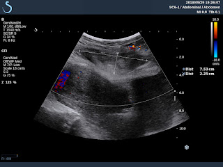
ULTRASOUND  DETECTED ONE MASS  IN HEPATIC BORDER ,  SIZE 5CM  ( US 1).


US  OBLIQUE SCANNING OF THIS MASS  IS THICKENING BORDER LIKE  AVOCADOS FRUID ( US 2).


US 3 : LIVER SCANNING OF THIS TUMOR  MADE BENDING   HILUS OF LIVER.


US 4 : ELASTOSCANNING OF THE WALL CODED  HARD PATTERN.

THERE WAS NO BLOOD INSIDE MASS AND ONE STONE  IN THE WALL OF THIS MASS  MADE THOUGHT IT GALLBLADDER.


CT SCAN WITH CE= CT1 : CROSSED SECTION  OF THIS
HYPODENSE MASS.


CT2 , CT3 :   FRONTAL VIEWS.


MRI OF ABDOMEN SHOWED THIS CYSTIC MASS, WITH ITS
VERY THICKENING WALL.


BLOOD TESTS = WBC:  1,9 K  WITH  8.9% NEUTRO,  CRP  96.8 ng/mL.
PRE OPERATIVE  DIAGNOSIS IS GALLBLADDER  ABSCESS WALLED OFF DUE TO STONE.
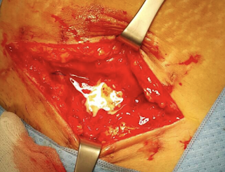
PHOTO OF MACROSCOPIC SPECIMEN IN   OPERATION FOR REMOVING GB ABSCESS.


CONCLUSION =  INFLAMMED GALLBLADDER NECROSIS BY STONE CAUSED OBSTRUCTION OF THE NECK OF GALL BLADDER.

Viewing all 624 articles
Browse latest View live