Quantcast
Channel: VIETNAMESE MEDIC ULTRASOUND
Viewing all 624 articles
Browse latest View live

CASE 499: RETINOBLASTOMA, Dr PHAN THANH HẢI, MEDIC MEDICAL CENTER, HCMC, VIETNAM.

$
0
0
BOY 3 YO  WITH RIGHT EYE  HAVING WHITE SPOT.
ULTRASOUND   B MODE OF  RIGHT EYE  DETECTED RETINE DETACHEMENT ( US1).

US B MODE   DETECTED  THE MASS TUMOR   INSIDE OCULUS [EYEBALL]  WITH CALCIFICATION (US 2, US 4).


MRI OF  THE TUMOR IN RIGHT OCULUS WITH  CALCIFICATION  NO  INVASION OUTSIDE.


CLINICAL ULTRASOUND AND MRI  SUSPECTED  RETINOBLASTOMA.
OPERATION  REPORT  IS  TUMOR LIMITED IN RIGHT OCULUS.  MICROSCOPIC RESULT IS  RETINOBLASTOMA.


CASE 500: BIG GIST TUMOR, Dr PHAN THANH HẢI, MEDIC MEDICAL CENTER, HCMC, VIETNAM.

$
0
0
Woman 65 yo  check -up  for hypertension.
US SCANNING of ABDOMEN   DETECTED   ONE MASS  SIZE OF 20CM X15 CM  AT THE  LEFT UPPER ABDOMEN QUADRAN,   SOLID STRUCTURE  AND MULTICYSTIC  HYPOVASCULAR (US1, US2).


X-RAYS of ABDOMEN WITH SP :THE MASS  DEPLACED AIR  GASTRIC  FUNDUS  AND LEFT COLON  ( X-RAY PICTURE).


ON MSCT CE   THIS MASS  CAUGHT VERY QUICK CE . IN CT 1( CROSSED SECTION),  CT 2:   THIS MASS IS  NEAR THE BORDER OF  GASTRIC GREAT CURVATURE , CT3 : FRONTAL VIEW,   CT4  SAGITAL VIEW .


GASTRO COLONOENDOSCOPY DID NOT DETECT ANY ABNORMALITIES  INTRALUMEN. 

BLOOD TEST IS NORMAL .
FOR THIS CASE  RADIOLOGIST  SUGGESTED GIST OF  GREAT OMENTUM.

Operation removed big tumor from the gastric wall .


CASE 501: BLADDER HERNIA to SCROTUM, Dr LÊ TỰ PHÚC, Dr PHAN THANH HẢI, MEDIC MEDICAL CENTER, HCMC, VIETNAM.

$
0
0
A 51-year-old man with pubic pain for 2 weeks with left scrotum swelling and
 increasing in size for 6 months. He has to  compress the scrotum by hand in order to empty the bladder everytime normal ending of micturition= two-stage micturition: the patient after a first spontaneous voiding, presses the mass and voids again. No other urinary symptoms. There was no history of nausea or vomiting.

Ultrasound scanning detected one sac containing of fluid on the left side of scrotum. This sac with thick wall continued upwards left pubic tubercle and getting smaller like a bird beak. This sac appears bigger when the patient did holding his breath (Valsalva maneuver) and disappered after being compression by hand to voiding.


Retrograde cystography CT revealed a left scrotal hernia with fluid density lesion continuous with left lateral bladder wall.


Diagnosis of left scrotal hernia with bladder as content was made.

Have you ever seen a case like that before and what do you think?

CASE 502: DIFFUSE SKIN TUMOR, Dr DƯƠNG NGỌC THÀNH, Dr LÝ HỮU ĐỨC, Dr PHAN THANH HẢI, MEDIC MEDICAL CENTER, HCMC, VIETNAM.

$
0
0


MAN 32 YO 2 YEARS AGO   BEGINNING AT THE HAND   AND FOOT  RISING SMALL NODULES AND HARD, SIZE 1-2 CM, COULORED  REED TO YELLOW   AND FULL BODY  APPEARED BUT NOT IN THE FACE. SEE FOTO   ( FOTO1  THE BACK ,  FOTO 2  THE FOOT,  FOTO 3   THE HAND,  FOTO 4  IN FOCUS NODULES,  FOTO 5  THE ACHILLE SKIN.


ULTRASOUND:   US 1=  THE NODULE 1,5 CM  UNDER SKIN   AND FATTY LAYER,   US 2 =  CDI  NO BLOOD TO THIS NODULE,  US 3  ACHILLE TENDON IS  NORMAL .


BLOOD TESTS MADE DIAGNOSING ARE DIABETES AND HYPERCHOLESTEROLEMIA AND RISING TRIGLYCERIDS.

BIOPSY  ( SEE MACRO).  


WAIT FOR  PATHOLOGY REPORT.
ONE DERMATOLOGIST SAYS T IS ERUPTIVE  XANTHOMATOSIS..

CASE 503: SUBUNGUAL GLOMUS TUMOR, Dr PHAN THANH HẢI, MEDIC MEDICAL CENTER, HCMC, VIETNAM.

$
0
0


WOMAN  46 YO  WITH  HISTORY  OF  5 YEARS AGO  PAIN AT  FIRST FINGER OF LEFT HAND, SWELLING AND BLEEDDING.
ULTRASOUND  OF THIS FINGER   WITH CDI    US 1   LONGITUDINAL SCANNING   DETECTED  ONE  MASS HYPOECHOIC BUT HYPERVASCULAR.


US 2: CROSSED-SECTION OF THIS MASS UNDER UNGUAL  
 HYPERVASCULAR  OF DISTAL FINGER.


MRI 2  =THIS MASS IS UNDER  UNGUAL   NO  INVASION OF THE BONE.


MRI 3 = CROSSED- SECTION OF THIS TUMOR IS  SUB UNGUAL WITHOUT EROSION OF THE BONE.


OPERATION  REMOVED THIS TUMOR.


 MICROSCOPIC RESULT IS  GLOMUS TUMOR.

CASE 504: PERIHEPATIC ABSCESS DUE TO FISHBONE, Dr PHAN THANH HẢI,Dr LÊ THỊ THANH THẢO, MEDIC MEDICAL CENTER, HCMC, VIETNAM

$
0
0


Woman 43 yo with  epigastric pain  has been treated as gastritis.
Ultrasound detected near  falciform ligamentun of liver  one abscess  with  foreign body long 3 cm  look like  fishbone  ( US1, US 2, US 3  SWE shows  it is very hard, US video).


CT scan no CE shows this fishbone is in a perihepatic abscess (CT1). 


One month later  the second CT examination with CE:  CT 2  shows  this abscess is  pulling abdominal wall out like a  hourglass [CT 2,  CT 3  sagital view].


Emergency operation of this abscess retrieved pus with a fishbone.


CASE 505: LEG ISCHEMIA, Dr PHAN THANH HẢI, MEDIC MEDICAL CENTER, HCMC, VIETNAM.

$
0
0

Man 70 yo in claudication with  pain at right leg and right foot changes in dark skin (photo).


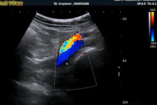
Vascular ultrasound:   
US 1=  normal left femoral artery and vein.


 US 2=  right femoral artery and vein in stenosis.


 US 3=  in middle of right  thigh cannot find out superficial artery.


 US 4= high flow of right dorsalis pedis artery.


Thermography  shows the right leg in hypothermia.


MSCT  angio of the leg arteries:  

CT 1=  big cysts in right/left kidneys and  abdominal aorta with sclerosis plaque.


CT 2=  right  superficial femoral artery is in obstruction. 


CT 3=  small anastomosis at the level of right thigh.


Diagnosis =  obstruction of right superficial femoral artery  with many sites of deep and superficial anastomosis of femoral artery.


DSA  shows  complete obstruction of right superficial femoral artery (DSA 1)


DSA 2 = small anastomosis.


Operation removed blood clot and revascularization of the right leg. Patient remains well recovery.


CASE 506: RENAL SINUS INFLAMMATORY PSEUDO- TUMOR, Dr PHAN THANH HẢI, Prof NGUYỄN TUẤN VINH, MEDIC MEDICAL CENTER, HCMC, VIETNAM.

$
0
0


Man 64 yo with clinical nocturia.
Renal ultrasound detected abnormal at right kidney.
US1 : Right big renal hilus and hyperechoic.


US 2:  Crossed-section of right kidney  shows hilus vascular compression.  


US 3:   Small size of right kidney  pelvis.


US 4:   In elastographic ultrasound, right renal hilus is hard.


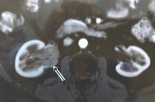
MRI of  abdomen.
MRI 1 = Right kidney in normal size, no hydronephrosis but renal hilus is covered by one mass  look like a tumor.


MRI 2 =  Mass of  solid structure covered the right renal hilus.


MRI 3 = In frontal view, right pelvic kidney is small by this mass compressed.


Endoscopic surgery=  right pelvic kidney is  normal  without tumor.


Laparoscopy  detected  the abnormal fatty mass covered the hilus kidney. Biopsy  no tumor detected.
Microscopic report is  inflammed fatty tissue of renal hilus, that is a pseudotumor.


CASE 507: URACHUS TUMOR, Dr PHAN THANH HẢI, Dr LÊ VĂN TÀI, MEDIC MEDICAL CENTER, HCMC, VIETNAM.

$
0
0


Man 54 yo with dysuria.
Abdominal  ultrasound at hypogastric region.
US 1=  longitudinal section over  suprapubic area, reveales one mass  from  abdomen wall and connected to urinary bladder wall  at urachus site. This mass is mixed structure with cystic and solid parts.


 US 2 = crossed section of this mass.


 US 3 = combination of 2 pictures scanning of intraurinary bladder that not detected any tumor.


MSCT  scan of  urinary system with CE.
CT 1:  crossed section  over urinary bladder.


CT 2:  sagittal scanning, this tumor is  related to urinary bladder wall and urachus,   calcification.


CT 3:  frontal view.


CT 4:  3D view of urinary system.


Radiologist report is  urachus tumor  looked like teratoma.
Operation to remove complete cystic tumor which  filled with mucus.


Wait for pathology report.
Conclusion: Ultrasound and CT make diagnostic of urachus cystic teratoma.

CASE 508: GALLBLADDER PSEUDOTUMOR, Dr PHAN THANH HẢI, MEDIC MEDICAL CENTER, HCMC, VIETNAM.

$
0
0


Man 26 yo with subhepatic pain post prandial for a long time.
Ultrasound of abdomen:
US 1=  intercostal scan,  liver is normal,  biliary tract no dilated, and  gallbladder (GB)  adheres in liver border  by 2 portions, one near the GB neck  filling by bile fluid and GB fundus covered by a solid mass with  size of 3 cm which is.well limited inside GB.


US 2=  Color Doppler (CDI):  no thickening of GB wall, no hypervascular in GB wall, and  no detected vascular supply for this mass. But no posterior shadowing of this mass with  little enhancement of the posterior wall.


US 3= the GB fundus is covered by this mass but the wall is intact. This mass has no motion.  


Sonologist suggested  a tumor of  GB  like GB adenomyomatosis.

MRI of the biliary tract.:
MRI 1=   the biliary tract has no stone and GB is filled by tumor at GB fundus.


MRI 2 = GB  has 3 portions, the middle portion  is hyperdense and adherent to liver. The GB wall is thickening like tumor and enhanced with gado.


MRI 3 = crossed section of the GB at middle portion, GB wall thickening and GB lumen is small.


Radiolodist report   is tumor of GB.
Laparocholecystectomy was performed.
Photo 1 =   the GB wall is well intact.


Photo 2,3 =  inside content of  material of black pigment like coffee waste. The GB wall is normal without tumor.


Pathology report is pigment sludge and inflammation of GB.

Conclusion=  Pseudotumor of GB  by intragallbladder sludge tumefaction. 

Reference  pdf  case report.


CASE 509: PANCREAS TUMOR, Dr PHAN THANH HẢI, MEDIC MEDICAL CENTER, HCMC, VIETNAM.

$
0
0


Woman  25 yo with history of  epigastric pain and  jaundice slowly for one month.
Ultrasound of liver:   big liver  with dilatation of the biliary system.  
US 1=  CBD  is in dilatation  # 2cm.


US 2 =  tumor from the head of pancreas and in extension to CBD.


US 3 =  head of pancreas tumor.


Ultrasound makes diagnostic that  pancreas head tumor moves to CBD.

MSCT  with CE  diagnoses again  pancreas head tumor.
CT 1 :  tumor looks like cystic pattern.


CT 2 : contrast enhancement in delay phase due to intratumoral bleeding.


CT 3 : vascular supply for this tumor.


CT  report by radiologist is pancreas tumor in invasion to CBD.  

Blood tests  =   CA19-9 = 4.96;  CEA= 0.56;   AFP=  0.3  
Summary of this case =  Young woman 25 yo has got  tumor of head pancreas in invasion to CBD.
Operation of Whipple is done.

MICROSCOPIC REPORT IT IS  SOLID PSEUDO PAPILLARY TUMOR  IT IS BENIGN  TUMOR OF PANCREAS 
 KNOWN AS   FRANKZ TUMOR.


COMMENT :  IN THIS CASE,  BLOOD TESTS CEA AND CA19-9 ARE NORMAL, THAT ARE  SAME IN REPORT  IN  REFERENCE CASE REPORT  BY WJG   2005.

REFERENCE =


CASE 510: MITTLELSCHMERZ SYNDROME, Dr PHAN THANH HẢI, MEDIC MEDICAL CENTER, HCMC, VIETNAM.

$
0
0


Woman 27 yo with  hypogastric pain. Ultrasound in emergency detected bleeding intra abdomen.
US 0=  fluid under liver area in  Morrison’s space.


US 1  = sagittal scanning at pelvis,  thickening of endometrium  and an amount of blood around pelvis.


US 2 =  transverse section at pelvis, right ovary too big  in comparison  to left one.


US 3 =  sagittal  mass at right  ovary.


Emergency blood test report  Hct 20%;  Hb 10 g/L; beta HCG  negative.


MRI 1 of abdomen detected  one mass at right  ovary.


MRI 2=  sagittal scanning of pelvis, retrouterus bleeding.


MRI 3=  frontal view of  right/left ovaries,  bleeding from right ovary.


Diagnosis is  bleeding from right ovary in Mittelschmerz syndrome.
Operation  removed 1000 ml blood clot  and right ovarian  rupture (photo) .


Mittelschmerz (German: "middle pain") is a medical term for "ovulation pain" or "midcycle pain". About 20% of women experience mittelschmerz, some every cycle, some intermittently.

CASE 511: LUNG CANCER,Dr PHAN THANH HẢI, MEDIC MEDICAL CENTER, HCMC, VIETNAM

$
0
0

Woman 60 yo with pain at left  lung after trauma. Chest x-rays detected rupture of 2 rib 
and mass at left lung ( see chest x-rays).


CT of  chest with CE


CT 1=   frontal view    


CT 2=  crossed section  


CT 3 = sagittal view


CT 4 with CE= very high CE enhancement.

Ultrasound at left lung  detected a tumor in invasion of the chest wall  (US1)   


US  2 = Color Doppler  shows  vascular supply to tumor   


Blood tests rised:  Cyfra 21-1 6.02 ( N=  3.3);   CEA=  7.13  ( N= 5).

Biopsy with CT guided is  carcinoma of the lung  adenocarcinoma 


Staging of this case  with  MSCT total body detected metastasis of the brain.


Conclusion:    Lung cancer detected accidentally by trauma,stage 4

CASE 512: TESTICULAR TUMOR, Dr PHAN THANH HẢI, MEDIC MEDICAL CENTER, HCMC, VIETNAM.

$
0
0


Man 43 yo  detected left testis swollen slowly.


Ultrasound  of scrotum  finds out  left  testis too big # 4 cm x 5 cm,   solid  hypoechoic looked like a hypovascular cyst.   
US 1=  crossed section,   hypoechoic in  comparison to right testis.


US 2 = hypovascular tumor


US 3 = elastoUS,  solid tumor # 25 kPa.  


MSCT with CE   this tumor is  quick CE enhanced with calcification.
CT 1 :  frontal view


CT 2 : sagittal view


CT 3:  sagittal view of left testis tumor


Blood tests=  beta HCG , AFP = normal levels; LDH=  is 313 (n :246)
Based on clinical , ultrasound, ACD, CT  diagnostic pre op is seminoma.
Operation  removed  tumor (see macro).


Microscopic result  is  seminoma.


DISCUSSION: Why does the testis tumor being hypoechoic looked like a cyst? 

CASE 513: KIDNEY TUMOR MIMICKING DROMEDARY HUMP, Dr PHAN THANH HẢI, Dr NGUYỄN MINH THIỀN, MEDIC MEDICAL CENTER, HCMC, VIETNAM.

$
0
0


Woman 41 yo with  righ kidney was detected abnormally  looked like dromedary  hump in general check up
Ultrasound  CDI:  US 1=  crossed section,  hypovascular pattern mass.


US 2=  longitudinal scan,  this mass liked a hump.


US 3 = elastoUS   inhomogeneous mass.


MSCT with CE, fast enhanced  contrast mass in CT 1, CT2.


MRI  with gado  shows  exophytic mass of the kidney border  (MRI 1, MRI 2).


MRI 3,4 : cystic structure  and bleeding inside.


Radiologis report is cystic tumor of righ kidney, BOSNIAK type 3 
Operation with  robot   removed  tumor in partial nephrectomy.


Operation of  this tumor  at righ kidney looked like  the seal.
 Specimen is cystic  septation,

Microspopic report is RCC.


Reference. Bosniak criteria.


CASE 514: IUD PENETRATING TO URINARY BLADDER, Dr PHAN THANH HAI, Dr JASMINE THANH XUAN, Dr NGUYEN MINH THIEN, Dr NGUYEN TUAN VINH, MEDIC MEDICAL CENTER, HCMC, VIETNAM.

$
0
0


Woman 49yo with pain after urinary miction. PAST HISTORY of BEING PUT T - SHAPE IUD 20 YEARS BEFORE.
ULTRASOUND of PELVIS  DETECTED BIG URINARY BLADDER STONE ( US1)

XRAY of PELVIS  SHOWs THIS STONE  WITH IUD  INSIDE   ( XRAY film).

ULTRASOUND   WITH CDI   FINDs OUT TWINKLING  ARTIFACT  WITH  COMET TAIL SIGN in GREEN AND RED ( US 2, US 3).


MSCT of  PELVIS  :
 CT1=  THE  METALIC IUD  INTRA UB WALL.

CT2  SAGITTAL VIEW .

CT3  FRONTAL VIEW : THE  IUD  INTRA UB WALL.

ENDOSCOPY DETECTED THE STONE IN VAULT OF UB.

CONCLUSION =  IUD  PENETRATING TO UB WALL AND FORMATION OF STONE.

CASE 515: VENTRAL HERNIA, Dr PHAN THANH HẢI, Dr VÕ THỊ THANH THẢO, MEDIC MEDICAL CENTER, HCMC, VIETNAM.

$
0
0


Woman  78 yo,  cesarian operation for  50 years, with, 2 days ago,  pain at left umbilical area  and everytime  coughing, a swollen mass appearing at the painful site.  

Ultrasound  of  this mass: 
US 1=  longitudinal scan at pelvis  near umbilicus, detected one subcutaneous mass. 


US 2 = left lateral scanning of middle line, shows  the tearing site of rectus muscle and a hernia goes out from it.


US 3 =  linear scanning of  this mass that was builded by great omentum and fluid.


US 4 = thickening of the bowel wall.


Emergency abdomen CT scanning:
CT  1:  crossed section  of  this mass  


CT 2 :  sagittal scanning of this mass


CT 3:  frontal section.


Emergency operation by  ventral hernia with past history of  cesarian operation 
detected  omentum and one bowel loop in ischemia but not necrosis yet.


Repaired this hernia by a mesh prosthesis. And timing is about in 4 hours from diagnostic to operation.

CASE 516: PANCREAS TUMOR, Dr PHAN THANH HAI Dr TRUONG DINH KHAI, MEDIC MEDICAL CENTER, HCMC, VIETNAM

$
0
0


Girl  10yo with  epigastric pain.
Ultrasound of abdomen detected  one mass of 8cm  at the body of pancreas, cystic  structure,  well bordered  ( US 1,  US 2, US 3),  no lymph node around.


MSCT with CE=   this mass is from the tail of pancreas,  inhomogeneous structure (CT1 CT 2, CT 3).


Blood tests  are normal.
Operation for resection of this tumor  [see macro].


Summary:  Girl 10 yo  with big mas at the pancreas,  structure is mixed solid and cystic,  the most common  is  solid pseudocystic papillary tumor.
Wait  for  histology.    

CASE 517: HCC WITH AFP LOW. Dr PHAN THANH HẢI, MEDIC MEDICAL CENTER, HCMC, VIETNAM.

$
0
0


MAN 52 YO with HBV INFECTED and   EPIGATRIC PAIN.   
ULTRASOUND CHECK UP DETECTED  LEFT LIVER TUMOR, SIZE 3.5 CM   HYPOECHOIC (US1,US2  )   BLOOD TETS: HBsAg POSITIVE, AFP 15ng/mL.


CT SCAN LIVER =  CT 1  NON CE , CT2, CT3   TRIPHASES CE , CT4 FRONTAL VIEW , CT5  SAGITTAL VIEW.

RADIOLOGIST REPORT IS  HCC.


MRI PRIMOVIST = MRI 1 DWI ,  MRI 2  T1, MRI 3 GADO INPUT,   MRI 4 GADO OUT,   MRI 5  FRONTAL VIEW.


BLOOD TEST AGAIN=  WAKO TEST  AFP-L3  RISING 16.4 %.


SUMMARY=  MAN 52 YO HBV INFECTED
WITH AFP NEGATIVE   BUT ULTRASOUND DETECTED TUMOR AND CT CE , MRI GADO   STUDYING THIS TUMOR.   WAKO TEST IS  POSITIVE  WITH L3 RISING. HOW ACCURATE DIAGNOSTIC OF   ALL FACILITIES?
OPERATION IN THE PLAN of LEFT HEPATECTOMY.
Robotic operation for  left hepatectomy.   See  macro  tumor with white area.

CASE 518: FINGER TUMORS, Dr PHAN THANH HẢI, Dr NGUYỄN NGHIỆP VĂN, Dr TRẦN THỊ THANH NGA, Dr LÊ THÔNG LƯU, MEDIC MEDICAL CENTER, HCMC, VIETNAM.

$
0
0
Woman 20yo with right hand  having many tumors at fingers,  slow growth, difficult movement of flexion ( see foto   (right hand  dorsal view:   tumor at first finger, finger number 3  and  number 5)  color skin  is changed,   soft in palpation and  no pain.


Ultrasound of  this tumor by linear probe 11 MHz : US 1:  tumor solid  hypoechoic, ellypsoid 3cm -2 cm, central necrosis ; crossed section ( US 2 ) with more vascular supplying and not fixed to bone.


Another tumor only in right hand not related to joint .
Clinical history :she was being treated in hospital as  hemangioma,   but  sonologist said  it is geant cell tumor of the tendon sheath.

X-Rays films of the right hand: No erosion of the bone.
Abnormal atrophic metatarsal number 4. 


Operation removed one small tumor at first finger.


Histology report is benign capillary hemangioma.


Viewing all 624 articles
Browse latest View live