Quantcast
Channel: VIETNAMESE MEDIC ULTRASOUND
Viewing all 624 articles
Browse latest View live

CASE 459 : CERVICAL LYMPH NODES, Dr PHAN THANH HẢI, MEDIC MEDICAL CENTER, HCMC, VIETNAM

$
0
0

Man 63 yo   detected  cervical  nodules  at right neck, that were in slow growth, no pain,  no fever,   no sore throat.
Clinical palpation this  lateral nodule of the neck  from SCM chain  continuous with subclavicular group
US scan  with 12 MHz probe= thyroid gland is normal


US1: many  small 1-2 cm hypoechoic nodes ,  round border.


US2:   big node =  round,  echo very poor ,  nonvascular inside.


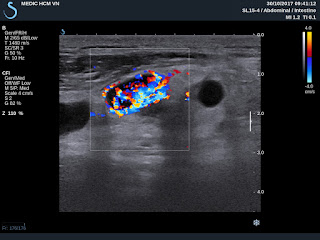
US3:  small node = very high vascular supply.

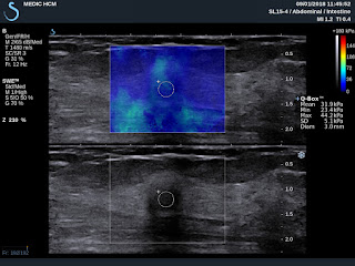
US4  elastoscan = very soft structure


And  the left neck is normal.


What is your suggestion for diagnosis?

CASE 460: THYROID CALCIFIED TUMOR , Dr PHAN THANH HẢI, Dr LÊ THANH LIÊM, Dr DƯƠNG NGỌC THÀNH, MEDIC MEDICAL CENTER, HCMC, VIETNAM

$
0
0


Man 52 yo, voice tone changes for  2 months, and  ENT doctor said vocal paralysis by endoscopy.
Ultrasound of the neck    
US1  left thyroid lobe normal

US2  right  lobe   covered by a big mass  4 cm  with strong  posterior shadowing  cannot see structure inside.


US3  near R/ CCA  small nodes with calcification #1cm.


US4  with convex probe ultrasound cannot se intra tumor by very strong calcification.


MTSC  non CE   


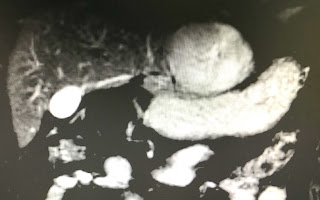
CT1: cross- section of the neck = mass is  very high calcification 

CT2 : cross- section=  calcification some lymph nodes near R/  CCA.


CT3 : frontal view  with CE=HU of this mass is  1,319 UI


CT4:   lymph node also has HU  1326UI.


CT5:  sagittal view   this mass  is  covered near the righ lobe of thyroid gland.


Blood test   TSH  is  0,041  T4  1,2   TG  97,42 (  n 3, 5-77)


Summary: For this case   clinical  ultrasound and CTt , blood test suggested   thyroid carcinoma   but report  of FNA  cytology is negative....

CASE 461: AXILLARY MAMMARY GLAND, Dr PHAN THANH HẢI, MEDIC MEDICAL CENTER, HCMC, VIETNAM

$
0
0

Woman  35 yo detected  at  right axiilary  a soft  and bigger mass. Clinical looked  like a lipoma.
Ultrasound  scan of this mass :  

US 1:  longitudinal scan=  subcutaneous hypoechoic mass,  size  4cm,  well bordered.   


US 2: CDI  hypovascular  pattern.


US 3:  elastoscan of this mass = 4.3 kPa,  like fatty tissue.


MSCT  non CE:

CT 1:  frontal view of  this mass  = subcutaneous, same density of fatty tissue.


CT 2 :  zooming of this mass  showed  structure  looked like a nipple of breast.


CT 3:   crossed-sectional view of  this mass = well bodered, not connected to the right breast.


CT 4:   sagittal view of  this mass=  separation to the right breast.


Radiologist reported  an axillary  mammary gland.
Operation  for removing  this mass     (see foto  specimen),  


surgeon reported it having  fatty  and hard tissue. Microscopic report is tissue of mammary gland.


Summary of  this case:    axiilary mammary gland  mimicking as a lipoma mass.

Article 0

$
0
0

Man 67 y o, emergency operation by acute necrosis of  gallbladder by stone one week ago, still pain at  Murphy area. WBC = 12k with neutro 90%,  CRP= 100ng/mL.
Abdomen ultrasound detected one mass # 5 cm  at the bed of gallbladder. Mass has got fluid content  and  white structures inside with very strong  shadowing and air in formatting an abscess.
No dilatation of the biliary system.
US 1: subhepatic abscess  with strong shadowing in abscess.


US  2: umbrella sign of  the shadowing.


US 3:  elatoscan  shows  this structure is very hard.


MSCT:   CT 1:  abscess with  air and fluid filling at the bed of gall bladder which had been  removed of GB.


              CT 2:  crossed-section view of this abscess:  inhomogenous structure  and air


              CT 3:   frontal view of the abscess.

   
Radiologist reported  textilloma in suspection.

Wait for reoperation. 

CASE 463: MADURA FOOT, Dr PHAN THANH HẢI, MEDIC MEDICAL CENTER, HCMC, VIETNAM

$
0
0

Man 25 yo, with  right  swollen foot   and  bleeding  after  small  trauma for  5 years.  Being treated  in many hospital  this foot  but  not better.  Biopsy 3 times in Cancer Center  with  report is chronic inflamation. (see foto 1,2).


Xray   shows  the  erosion of metatarsal bones ( xray)


Ultrasound  reported   edema with  hypervascular  soft tissue of the foot 


Biopsy of this  tumor again.

Waiting for result.

CASE 464: ANTERIOR MEDIASTINAL TUMOR, Dr PHAN THANH HẢI, MEDIC MEDICAL CENTER, HCMC, VIETNAM.

$
0
0


Female patient 62 yo, cardiologist send to CT scan for coronary artery  but in same time CT scanning detected one 3 cm mass  at the superior anterior madiastinal area.., well bordered.

CT1:  crossed section  non CE this mass has HU  = 54.


CT 2 :  after CE late phase HU  = 73.


CT 3 :  sagittal section, this mass is  ovoid  form at the  anterior mediastinal area.


CT 4 :  frontal view,  this mass is near aortic ascending.


CT 5 :  CE  arterial  phase,  this mass is late enhanced  and cystic formation.


Blood test is normal, and negative all cancer markers.


Radiologist suggested  thymoma. What do you think about ?

CASE 465: LUNG with LOFFLER SYNDROME, Dr PHAN THANH HAI. MEDIC MEDICAL CENTER, HCMC VIETNAM

$
0
0

Woman  45 yo with  fever and cough. Chest XRays detected many  white spots  like balloon both site the lung.


Radiologist suggested diffuse lung metastasis.
Blood tests=  wbc rise 11,27 k  eosi 20,7%  IgE 1779ui/ml
Toxocara sp  positive  with od 1,809 and  all cancer marker are negative.
CT scan  with CE many opacification of  peripheral lung  booth site( CT1a/b)


CT scan the lung  with CE   the  lesion is small  regression.


Ultrasound of thorax  with small light 


After one week treatment  no fever  no cough.   Chest  Xrays  is clear.


Summary:   By the clinical, blood tests, chest XRays,  diagnosis  as  Loffler syndrome  of the lung  was made for the case.

CASE 466:CHILDREN HEEL PAIN : A SEVER'S DISEASE CASE , Dr PHAN THANH HẢI PHƯỢNG, MEDIC MEDICAL CENTER, HMC, VIETNAM

$
0
0
9 years old male patient,  with chief complain of pain in both heels, which worsen by physical activity such as walking, running.
Physical examination: generally normal, Squeeze test (+) on right side.

 X-ray examination and ultrasound was performed.

On ultrasound plantar fascia is normal. Note: the anechoic region between calcaneous is not fluid (which can indirectly suggest fascilitis in case of adult) but infact the normal apophysis (growth plate). 


Achilles tendon is normal and remains continous fibrous echotexture (US 2), again, the rough bone surface with anechoic shown normal apophysis.

Normal distance to apophysis in both sides, no dislocation, no avulsion.


X-rays examination of both 2 heel  are normal.


Physician suggest Sever's disease, and patient was advided rest and proper physical activity and shoes fitting.

Conclusion:
Sever's disease, the most common cause of children heel pain, known as calcaneal apophysitis is an inflammation of growth plate in heel of growing children.  Diagnosis usually bases on clinical,  and X-rays is normal. Ultrasound is suitable diagnostic tool while X-ray examination is only helpful when an ossification center of apophysis exist. Ultrasound  helps ruling out muscle strain, detect edema, lytic and avulsion.

CASE 467: SUBMANDIBULAR MASS, Dr PHẠM THỊ THANH XUÂN, Dr PHAN THANH HẢI, MEDIC MEDICAL CENTER, HCMC VIETNAM

$
0
0

Man 41 yo,   5 years ago  detected one mass at submandibular region, slow growth. Clinical palpation no pain,  soft, and without  trouble of  eating.


Ultrasound:
US 1=  longitudinal  scan with  curve probe 3.5 MHz :   ovoid mass clear border, hypoechoic with  no vascular signals inside.


US 2 = scanning with linear probe 12MHz=   inhomogeneous structure with many  black spots,    size # 0.5 cm.


US 3 =  elastoscanning of   this mass disclosed a cyst  with  many spots  hardening;  like pomegranate fruit.


CT scan with CE=   it is a cyst,  well bordered, .CT1, 2 , 3 with 3 sections of  this mass,    radiologist said  teratoma.


Operation for  remove this cyst with  content like  yellow milk   typical of  sebaceous cyst  (epidermoid cyst).


 MICROSCOPIC REPORT  IS EPIDERMOID CYST,   BENIGN TUMOR.


CASE 468: ADRENAL GLAND TUMOR, Dr PHAN THANH HẢI, Dr NGUYỄN PHỨƠC TÒAN, Dr VĨNH TUẤN, MEDIC MEDICAL CENTER, HCMC, VIETNAM

$
0
0

Man 26yo,  ultrasound of abdomen for screening  incidentally detected  one mass of 5 cm at the right adrenal gland area.
US1= longitudinal scanning of  this tumor at upper area of right kidney, well bordered.

  
US2=  CDI of  this mass pulled down right kidney.


US3=  crossed section  of this mass is well bordered  under liver near IVC.


US4=  very  small vascular signals  in mass.


  • US5=   elastoscanning of this mass: very hard  32 kPa in comparison to  liver = 9.3 kPa.


Sonologist reported solid adrenal tumor for this mass.

MSCT revealed:
CT 1= this mass is  hypodense like a cyst.


MSCT with CE, CT 2=  this mass is very low enhancement.


CT 3=  crossed section and sagittal scanning like a cyst of adrenal tumor.


Blood tests :  no abnornal  of cortico-medullary  adrenal function.
Pre-op   suggestion of surgeon is cyst of adrenal gland.
OPERATION REMOVED THIS TUMOR COMPLETLY.  MACROSCOPIC SPECIMEN WAS  WHITE AND HARD STRUCTURE, SECTION SURFACE SWELLED UP.
MICROSPIC REPORT IS  PARAGANGLION NEUROMA, BENIGN TUMOR.


CASE 469: T CELL LYMPHOMA OF SUBMANDIBULAR NODES, Dr PHAN THANH HAI, MEDIC MEDICAL CENTER, HCMC

$
0
0

Man 30 yo, detected 3 months ago submandibular nodules, subcutaneous neck , no fever.


Ultrasound of the neck
US 1: normal thyroid , and subcutaneous nodule is round, black like a cyst .


US 2:  Lymph nodes have the hilus.


US 3:  CDI  Hypervascular with the vascular tree.


US 4: MP4  VIDEO


US 5:  PW DOPPLER  RI  0.56,  PSV  46cm/S 


US 6:  ELASTOSCAN kPa 0.2 


Clinical suspected  lymph node lymphoma.
Blood test   EBV IgG positive   118.5 UI/mL  IgM  negative 

BETAMICROGLOBULINE   2264UI (N  1412)
Biopsy and  histobiochemistry report is   T-CELL LYMPHOMA.


CASE 470: TROISIER-VIRCHOW NODES, Dr PHAN THANH HAI, MEDIC MEDICAL CENTER, HCMC, VIETNAM.

$
0
0
MAN 70 YO  COUGH . NO FEVER. CHEST CT   DETECTION  IS LEFT LUNG  LESION  WITH WIDERING OF MEDIASTINUM.
(CT1/ CT2:   SUPRA LEFT CLAVICULAR  MASS SUSPECTED LYMPH NODES.


ULTRASOUND  of SUPRA LEFT CLAVICULAR  AREA IS  MULTIPLE LYMPH NODES.
US1=LYMPH NODES ROUND 2CM, LOSS HILUS, HYPOECHOIC,  HYPOVASCULAR.


US 2 =THE VASCULAR SUPPLY IS  INTER-NODES  LIKE A RING.


US 3= ELASTOSCAN of THIS NODES WITH CENTRAL 23 kPa.

ULTRASOUND EXAMINATION REPORT IS METASTASIS  LYMPH NODES   THAT WERE TROISIER-VIRCHOW NODES.
WAIT FOR  BIOPSY.
MICROPATHOLOGY REPORT WITH  IMMUNOHISTOCHEMISTRY  IS   NEURO ENDOCRINO-
CARCINOMA METASTASIS TO  TROISIER -VIRCHOW NODE.


REFERENCE :  ANATOMY TROISIER NODE

                         PICTURE OF  DR TROISIER --PROF VIRCHOW.


CASE 471: MESENTERIC LYMPHOMA, Dr PHAN THANH HẢI, MEDIC MEDICAL CENTER, HCMC, VIETNAM.

$
0
0

Man 70yo  with  epigastric pain. Ultrasound of abdomen detected  one hypoechoic mass like a cyst # 4.9cm near SMA.
US 1=   longitudinal scan at epigastric area.

US 2 = CDI:   this mass  is near SMA.

US  3 = crossed scanning= this mass  is placed over the head of pancreas in relation  with  celiac artery and vein.

US 4  some  small lymph nodes near the big mass .


MSCT CE=
CT  1 : crossed section  this mass  over the artery and vein.


CT 2:  the pancreas is normal.

   
CT 3:  frontal section of this intramesenteric mass near  SMA and vein   with some  lymph nodes in mesentery.


Endoscopy gastric and colon are normal, blood test and all cancer markers are normal 
What is your  suggestion for diagnosis?


CASE 472: INCIDENTAL BREAST TUMOR , Dr PHAN THANH HẢI, MEDIC MDICAL CENTER, HCMC, VIETNAM

$
0
0


WOMAN 58YO   FOR SCREENING  TO DO WB MSCT. 
RADIOLOGIST REPORTED ONE MASS AT  RIGHT  BREAST  SIZE# 1.3 CM.(  CT1  FRONTAL VIEW, CT2  AXILLARY AREA).


ULTRASOUND POST CT VERIFIED THIS MASS  WITH  SIZE  # 2 CM   VERY STRONG  SHADOWING, AND HYPERVASCULAR.


US1,  US2  ELASTO  IS  31.9 kPa .
US3 , AXILLARY NODE    SIZE 1.5CM WITH HILUS 
US4 ,  CDI  = VASCULAR  HILUS DEFORMATION.


CORE BIOPSY  RESULT IS  BREAST CANCER, STAGE T2 N2.


CASE 473: SCIATIC NERVE TUMOR, Dr PHAN THANH HẢI, MEDIC MEDICAL CENTER, HCMC, VIETNAM.

$
0
0

Woman 59yo  detected  one mass  at right buttock that is in slow growth and no painfull.
Ultrasound scan
US 1= longitudinal scan of  this mass with size 6.7 cm,  ovoid, hypoechoic  in gluteus maximus muscle   near sciatic nerve  like a size of a mango.


US 2 = crossed section of this tumor is well bordered and hypovascular pattern. 

US 3 = elasoscanning of  this mass is inhomogeneous  structure.


MRI scan


MRI 1   crossed section   this mass is   well bordered  in a muscle   
MRI 2   frontal view this mas is  bordered of right sciatic nerve.
MRI 3   relation of this mass and right sciatic nerve.


Core  biopsy is done and histology report is   neuroma.


CASE 474: LEFT LIVER LOBE TUMOR, Dr PHAN THANH HẢI, MEDIC MEDICAL CENTER, HCMC, VIETNAM

$
0
0

Man  52 yo  with chronic hepatitis  HBV,    follow up  every 6 months  and  AFP, HBV -DNA  negative. But  ultrasound   detected  big tumor  6 cm in diameter at left lobe of liver.
US  1:  transverse scan  this tumor is well bordered,   hypoechoic pattern.
US 2:  longitudinal scan  at tumor site.
US 3:   color Doppler=   the vascular supply to tumor is from left  liver.


MRI  with Primovist   uptaked  and washed out  as a HCC.( MRI 1, 2, 3, 4).


Blood test  Wako  only DCP  raised 91 UI.


Summary:  in case chronic hepatitis HBV   ultrasound detected  big tumor in liver, Wako test  only DCP raised.

What is your suggestion for diagnosis?

CASE 475: INCIDENTAL THYROID CARCINOMA ON CT, Dr PHAN THANH HẢI, MEDIC MEDICAL CENTER, HCMC, VIETNAM.

$
0
0

Man 55 yo for check- up  MSCT  total body and blood tests.

Radiologist report  detected one mass # 1.6 cm at right lobe  of thyroid gland,  with  HU 91 UI  in comparison to  the thyroid tissue background =121UI ( CT1  CT2  CT3).


Blood test is normal thyroid function.

Ultrasound scanning is second look:

US 1:  crossed-section  of the hypoechoic  focal  lesion, well-bordered #  1.5 cm.
US 2:  longitudinal scanning of  the mass is 1. 7 cm, hypoechoic pattern,  with 
US 3:  CDI = hypovascular  mass,  no lymph node in the neck.


This mass is  in TI-RADS 3, need  FNAC.
FNAC report suspected  papillary carcinoma ( PTC).


Operation is done for subtotal thyroidectomy (see macro 1,2).


POST OP  PATHOLOGY REPORT IT IS PTC [PAPILLARY THYROID CARCINOMA].


REFERENCE:


CASE 476: ABDOMINAL WALL TUMOR, Dr PHAN THANH HẢI, Dr VÕ NGUYỄN THỤC QUYÊN, MEDIC MEDICAL CENTER, HCMC, VIETNAM.

$
0
0


Woman 60 yo in general check- up,  ultrasound of abdomen detected one mass  6x4 cm  at RLQA.

US 1:  longitudinal scanning over pelvic region  disclosed one mass near urinary bladder.

US 2:  crossed –section  of  hypoechoic mass,  well bordered,  no change position when patient moves  to lateral decubitus.


 US 3:  CDI = hypovascular mass. The vascular supply of tumor from the abdomen wall.


 US 4 : linear probe presented  the tumor and abdominal wall.


US 5:  elastoscanning of   inhomogeneous structure of tumor.

Blood test is normal
MRI  with gado: 3 position scannings of  this tumor . Radiologist says  mesenteric tumor.


Laparoscopic operation: This tumor is extra peritoneum.  Macro view looked like Desmoid tumor.


CASE 477: LEFT EXOPTHALMIC EYE, Dr PHAN THANH HAI, MEDIC MEDICAL CENTER, HCMC, VIETNAM

$
0
0


Man 52 yo with  cough and pain at left eye, protrusion the orbis and edema the cornea for one week (photo).

MRI of the eye and or brain.


MRI 1=   frontal view  of the left exopthalmic eye.
MRI 2=  frontal section of the left orbis.  edema of the  intra orbis muscles,
MRI 3=   crossed section of left orbis  : left superior opthalmic vein dilated.  
MRI 4=   the muscles in orbis are edema  and  cavernous sinus  is not  abnormal.
For  make  sure diagnostic  DSA was done that  detected  A-V fistula at  cavernous sinus.
DSA  with dilated opthalmic vein=   DSA1  putting of the coil,   DSA 2 after treatment. Coil embolisation is  spectacular reduction clinical sign  (photo 2).


The left eye returns  near normal 24 hrs after treatment.


Conclusion:    Basis clinical signs of MRI and DSA   can make diagnosis  and spectacular  treatment. success. 

Reference:  Anatomy of eye circulation.


CASE 478: POPEYE’ SIGN of BICEPS MUSCLE, Dr PHAN THANH HẢI, MEDIC MEDICAL CENTER, HCMC, VIETNAM.

$
0
0


Man 58 yo playing tennis got pain in left upper arm.  Clinical  detected left biceps muscle protrusion  in contraction and painful. ( see photo 1 relaxed position,   photo 2  flexion position).


Ultrasound of biceps muscle  =

US 1 :  longitudinal scanning represented   rupture at the upper head of biceps muscle.


US 2 :  crossed section disclosed  echo poor pattern due to hematoma.  


US 3 : crossed section at middle part of biceps muscle  showed  hyperechoic pattern  by contraction.


MRI  made sure that rupture of upper part of biceps muscle.


Operation for repairing the ruptured muscle.   

Conclusion:  Orthopedic pathology due to sports named  Popeye' sign. 

Reference:   Anatomy of biceps tendon  and  Popeye’ sign. 


Viewing all 624 articles
Browse latest View live