Quantcast
Channel: VIETNAMESE MEDIC ULTRASOUND
Viewing all 624 articles
Browse latest View live

CASE 440: MULTIPLE TUMORS of the LEG, Dr PHAN THANH HẢI, MEDIC MEDICAL CENTER, HCMC, VIETNAM

$
0
0

Man 20 yo with  history of 10 years ago having many small subcutaneous  tumors on left  foot ,  size of 2 cm, no pain. And now he detected another nodule near his left knee (see photo1, photo 2).


 It  is soft in palpation, no pain,  compressible and reexpansion after releasing it.
Ultrasound examination of  this tumor showed  tumor belonging to sapheneous vein while  deep vein is normal.


MRI reported that tumor of superficial vein of left foot.


Biopsy of this small  tumor. Microscopic and histoimmuno staining is angioleiomyoma.


CASE 441: CHERRY-LIKED SKIN TUMOR, Dr PHAN THANH HẢI, MEDIC MEDICAL CENTER, HCMC, VIETNAM

$
0
0

Lady 17 yo  with tumor  on her left shoulder   which was  detected for  2 weeks,  size of 3 cm, red and soft,  no pain ( see photo 1,2).


On ultrasound examination,  this tumor  came from  skin layer, inhomogenous with solid part in the root and calcification, while upper part is fluid (US 1, US 2). On CDI, tumor  has hyper vascular pattern like an octopus.


What is your diagnosis ?

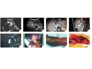
CASE 442: BILOBATE GALLBLADDER: Dr PHAN THANH HẢI - Dr LÊ THANH LIÊM, MEDIC MEDICAL CENTER, HCMC, VIETNAM. .

$
0
0

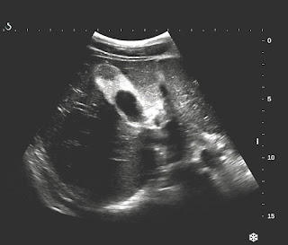
Male 18yo  with recurrent right subcostal pain.  
Ultrasound of abdomen detected abnormal gallbladder, size of 5 cm, bilobe, thickening wall of GB fundus  with more  twinkling artifact pattern on Doppler examination.


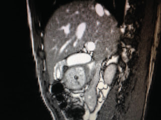
MRI of liver and biliary confirmed the morphology of gallbladder, its shape is bilobe and abnormal signal of GB fundus.


Radiologist report is gallbladder adenomyomatosis.


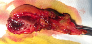
Operation laparoscopy removed the bilobate gallbladder.
Microscopic report is adenomyomatosis  of gallbladder.


Reference : US, CT , MRI diagnosis of   adenomyomatosis gall bladder


NHÂN CA 312 ĐƯỢC ĐĂNG TRÊN ULTRASOUND (UK) 2017, Vol 25(2)

$
0
0
CASE 312 : Fetal Body Hyperflexion { PENA-SHOKEIR phenotype} published on ULTRASOUND 2017, Vol 25(2) sagepub.co.uk/journal SAGE DOI: 10.1177/1742271X16688235

case 312 VUD

Xem fulltext theo link
https://app.luminpdf.com/viewer/ihfxeZZRFjjr8zsi3


CASE 443: DUODENAL TUMOR, Dr PHAN THANH HẢI, Dr LÊ ĐÌNH TÍN, MEDIC MEDICAL CENTER, HCMC, VIETNAM

$
0
0
Man 54yo with epigastric pain  crisis  after eating.
Ultrasound of abdomen  detected  tumor  at duodenum  D3, size of  3 cm,  hypoechoic (US 1),  and CDI  showed  hypervascular tumor  (US 2, US 3, US 4).


Gastrograph  with barium meal detected  filling defect  at  duodenum D 3 ( X-ray1, X-ray 2).


MSCT with CE:revealed  this tumor  very quick and high contrast enhanced (CT 1, 2, 3, 4).


Normal blood tests, and  nothing abnormal detected on endoscopy.

What is  your suggestion for diagnosis ?

CASE 444: FEVER in PREGNANCY, Dr PHAN THANH HẢI, MEDIC MEDICAL CENTER, HCMC, VIETNAM

$
0
0


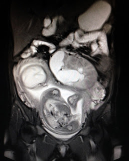
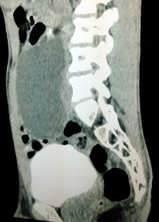
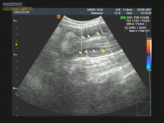
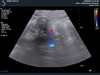
Woman 23 yo, 28 weeks primigravida with 5 days fever and abdominal pain in  periodic crisis.
WBC=  27 k  with neutro 97% ;  CRP= 342 mg/L,  normal urine test . And clinical suspected  appendicitis.
Ultrasound of  abdomen reported 29w pregnancy and  normal status of fetus. One cystic mass retro-uterus  near left pelvis  iliac artery,  suspected  left kidney  ectopic pelvis with hydro-pyonephrosis while  right kidney is normal ( US 1, US 2, US 3, US 4).


MRI  reported  left kidney hydronephrosis due to ureter obstruction.


Emergency cystoendoscopy was done for putting  stent  to release pus out from left kidney.


Conclusion=Pregnancy with  fever due to left pyohydronephrotic kidney as obstruction of ureter.

CASE 445: PEDUNCULATED LIVER TUMOR, Dr PHAN THANH HẢI, MEDIC MEDICAL CENTER, HCMC, VIETNAM

$
0
0
Woman 33 yo  with epigastric pain  has been treated as  gastritis.
Ultrasound of  abdomen detected  one mass near  the liver border having hypoechoic peduncule from liver,  and changing position with  respiration movement (US1). There is vessel from the liver for peduncule of tumor (US ). In  cross-sectionnal scanning,  this tumor represented its well bordered, solid, hypervascular structure (US 3)


MSCT with CE detected this tumor in  connecting with  the liver by a long peduncule ( CT 1,sagital view) and in  frontal view, this mass is  nearby the  ligamentum falciformis (CT2).


CT 3: cross sectionnal of  tumor = well contrasted enhancement.
CT 4: vascular supplying for this tumor is a branch from  left gastric artery and  another one from liver.


What is  your  suggestion of diagnosing for this tumor?

CASE 446: KILT SYNDROME, Dr PHAN THANH HẢI, MEDIC MEDICAL CENTER, HCMC, VIETNAM

$
0
0


Man 35 yo,  hematuria repeated  many times with  many  dilated subcutaneous veins on the abdomen wall [photo].


US of  kidneys   showed  righ and left kidney looked like hydronephrosis. But cannot detected the cause ( US 1, US 2)   US 3  =  CDI of  renal artery;  US 4 = spectral Doppler of pyramid arteries; US 5 = varicosis in urinary bladder wall.


CT of kidney without CE ( CT 1)=   HU of pelvis kidney  = 8 UI
CT 2  with CE ,  CT 3: secretion phase; CT 4= small ureter on R and L sides.


 MRI 1, MRI 2= kidney non CE   showed pelvis kidneys dilated and fluid collection around  2 kidneys.


Ultrasound of the leg = dilated chronic  deep vein thrombosis.

Conclusion= absence of IVC  suprarenal with DVT of  the legs, itis  K I L T syndrome.


CASE 447: RETROPERITONEUM CYST, Dr PHAN THANH HẢI, MEDIC MEDICAL CENTER, HCMC, VIETNAM

$
0
0

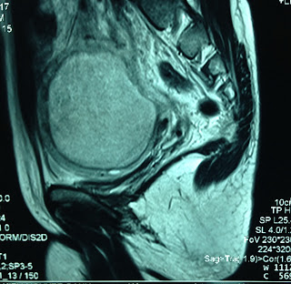
Man 20 yo with abdomen distention at  right subcostal region,  no pain  no fever.
Ultrasound of abdomen detected  one big mass in retroperitoneum, multiseptation and  multiloculated in connection with troubled fluid inside.
US 1=  longitudinal scan  over right kidney.
US 2 = crossed sectionnal scan over aorta.
US 3 = longitudinal  scan over spine.
US 4 = longitudinal  scan at  pelvis.


MSCT of  abdomen with CE:


CT 1 = sagital scan of  cystic mass  retroperitoneum.
CT 2 = frontal scan  of this mass  is cystic homogeneous.
CT 3 = frontal view of  this mass  and  gastrointestinal mass arround.
CT 4 =  aorta  no deplacement.
Normal blood test and  all biocancer markers.
What is  your  suggestion for diagnosis?


CASE 448 : FEVER and ASCITES, Dr PHAN THANH HẢI, MEDIC MEDICAL CENTER, HCMC,VIETNAM

$
0
0

Woman 40 yo with one week  fever,  abdomen pain  and distention..
Clinical examination ruled out  surgical needs , chest  X-ray is normal.


Ultrasound of  abdomen = liver, biliary system,  kidney are normal, huge amount of  ascites volume with cloudy fluid  (us1 , us2, us3 pelvis  us4 ovary).


CT scan of abdomen  =  No tumor detection [ ct1,frontal view, ct2, ct3  cross section].


Blood tests =  WBC  15k with 13,3 k neutro, CRP= 25.9,  amylase, CEA , CA 125  are  normal level.  But Widal test is  positive th;1/320
Ascites punction= yellow  clear,   analysis =  ADA= 19.5 ng/mL,  CA125 :396 UI/mL, CEA: 0,8UI,  albumine =3.9 mg/mL


After 2 weeks treated with antibiotics;   response is good,   no fever  but ascites  is  distention after  aspiration  many times.
Summary=   Fever with typhoid  but ascites  still  persistent after one month.

What we can do for this case?


CASE 449: LIVER TUMOR, Dr PHAN THANH HẢI, MEDIC MEDICAL CENTER, HCMC, VIETNAM

$
0
0


Man 77yo, in  general check up  by ultrasound  detected  one round mass 5 cm in  segment 6 of liver with Left  kidney cyst and big prostate  (US 1, US 2, US 3, US 4).


MSCT of liver with CE=   showed this liver tumor  well bordered, quick CE   and  delay wash out CE that looked like HCC by radiologist  (CT1, CT 2, CT 3, CT 4).


Blood tests = HCV , HBV non detected. Wako test =  3 negative.


In MSCTA, vascular supply of the tumor comes from left gastric artery.


Operation laparotomy  removed this tumor  that macroscopic feature likes liver normal tissue of liver.


CASE 450: PID with PELVIC MASS, Dr PHAN THANH HẢI, MEDIC MEDICAL CENTER, HCMC, VIETNAM

$
0
0
Woman  34 yo with  onset 2 weeks ago,  fever  and pain at pelvic region.
Ultrasound  first at an obgyn hospital  says  ovary cyst or endometriosis. But medical  management  with antibiotis failed in clearing fever.


(US 1: scanning  after 2wks,  uterus and cystic mass # 10cm with very thickening wall; US 2 : on CDI;  US 3:  intracystic mass detecting one structure like a bridge: US 4:  ascites intra RLAQ;  US 5:  this mass was scanned with linear 12 MHz probe. 
MRI of pelvis with gado=   this cystic mass is  in left ovary  with  the wall very thick, and  black spot intra mass  unknown  original,   but radiologist  suspected an ovary cancer.


Blood tests=   WBC  12k with 70% neutron,  Plt= 515,  CA 125  rising  100 UI.

What is your  diagnosis for the case?  


CASE 451: CARDIAC MYXOMA, Dr PHAN THANH HẢI Dr NGUYỄN TUẤN VŨ, MEDIC MEDICAL CENTER, HCMC, VIETNAM.

$
0
0

Woman 33 yo, with  dypsnea progressing for one year but being normal chest X-Rays , EKG and abdomen ultrasound.   

  
Cardiac ultrasound  detected  one mass 5 cm intra left atrium  covering  near all cavity space ( US 1, US 2, US 3). 


And  echocardiologist says  cardiac myxoma.
CTA  of cardiology  non CE showed  this round mass  related to  left atrium, and radiologist confirmed  myxoma in left atrium.


Emergency heart operation was done.
OPERATION  REMOVED BIG TUMOR 5 CM  WITH  STRUCTURE LIKE  MYXOMA.


MICROSCOPIC REPORT IS MYXOMA.

CASE 452: MESENTERIC TUMOR , Dr PHAN THANH HẢI, MEDIC MEDICAL CENTER, HCMC, VIETNAM.

$
0
0
Woman 65 yo with  anorexia,  weigh loss # 5 kg in 3 months.
Ultrasound of abdomen for general check-updetected  one round mass, mobile  at  RLQ
US 1 :  hypoechoic homogeneous  mass looked like a cyst,  size 3 cm, round border  with  low posterior enhancement,  intra mesenteric situation.


US 2 : CDI   vascular  around this mass not branching inside this mass   
US 3: CDI with another section, no vascular in the mass
US 4 scanning by linear 14 MHz  probe:  this mas  like a cyst.
US 5 :  elastoscan of this mass  mean  9kPascal.

MSCT with CE=


CT 1:  crossed section of this mass very quick and  high contrast enhanced,  

CT 2:  frontal section of  this mass = intra mesentery

CT 3:  sagittal section of this mas:  near  anterior wall of abdomen  

Blood tests= normal  CBC, negative all cancer markers.


What is your suggestion of diagnosis?

CASE 453: SMALL BOWEL MELANOMA, Dr PHAN THANH HẢI, MEDIC MEDICAL CENTER, HCMC, VIETNAM

$
0
0

Man 56 yo with acute abdomen pain,  vomitting, and blackwish stool. Clinical examination was oriented to 4tday bowel occlusion.


Abdomen US scan in emergency  detected  dilated bowel  with  crossed sectional view  presented typical oignon sign  of intussusception  ( US 1,   crossed section;  US 2,  longitudinal  scan.    With linear probe, US 3, CDI examination;  US 4,  multilayer of  intussuscipiens [boudin].


MSCT  with CE of abdomen =   


CT 1: bowel dilatation  due to  bowel obstruction
CT 2 : mass  with  multilayer of small bowel wall.
CT 3 :  intussusception with target sign or pseudokidney sign
CT4 :  sagittal view of the abdomen
Lab test is normal.


What is your  emergent diagnosis for the case?

CASE 454: IVC STENOSIS, Dr PHAN THANH HẢI, Dr TRỊNH DUY TRANG, MEDIC MEDICAL CENTER, HCMC, VIETNAM.

$
0
0

Woman 30 yo with 3 times of miscarriage, she came to MEDIC for a check- up (foto  subcutaneous veins).


Ultrasound of abdomen and pelvis:  normal uterus size.


US 1 = big liver caudate lobe 
US 2 = IVC  stenosis at  upper portion of liver
US 3 = crossed section of IVC no flow with hepatic vein.

MSCE with CE:


CT 1=  normal uterus structure.  CT2  = IVC  contrast filling  short portion cannot go upper to liver portion.  CT3 =  crossed section of  dilated subcutaneous abdominal veins. CT4 = crossed section= IVC no contrast in liver portion and abnormal late phase of liver vein, 
CT 5  = surface abdomen skin.


TREATMENT  ANGIOINTERVENTION;   DILATATION of STENOSIS AND STENTING.


Summary  =  IVC abnormal  stenosis near  diaphragm and  many venous collateral returning ways. 

REFERENCE:


CASE 455: FINGER TUMOR, Dr PHAN THANH HAI Dr TRUONG TRI HUU, MEDIC MEDICAL CENTER, HCMC, VIETNAM

$
0
0

Woman 54 yo,  4 months  ago  detected  one mass, at 4th finger of right hand,  slow growth,  no pain,  no disturbing movement.of this finger  (see  photo1, 2).


X-Rays  of AP and lateral views of 4th finger  = bone is  normal but  periosteum changing this mass to a soft tissue tumor ( xrays 1, 2).


Ultrasound   scan of this mass is hypoechoic like a cyst of lateral finger, from the tendon,  size 3 cm of length (US 1).
US 2  CDI of  vascular supplying arround this tumor means  a solid tumor
US 3  crossed section  of the arround vascular tumor.
US 4 mass is soft  on elastoscanning , arround 30 kPa.


What is your  suggestion  for diagnosing the finger mass?    

CASE 456 : LEG GAS GANGRENE, Dr PHAN THANH HẢI, MEDIC MEDICAL CENTER, HCMC, VIETNAM

$
0
0


Man 41 yo,with  history of liver cirhosis and ascites. 3 days fever and pain at left swollen leg.
      [Foto1]. 


    Clinical suspected lower limb DVT
Ultrasound  rouled out DVT ( US 1=artery and vein at left inguinal), US 2   fluid collecting 
       between muscle quadriceps; US 3  scanning at calf  detected intramuscular  air
        US 4 air and  level.


MSCT non CE detected   air in calf muscles
CT 1  crossed section;  CT 2  sagittal scanning   


Blood test, WBC  no rising;    blood culture  detected  gram  negative bacillus, 
 24 hours after admission hospital patient died   with  bullous leg [Foto2]


CASE 457: AML KIDNEY TUMOR, Dr PHAN THANH HẢI, Dr NGUYỄN HOÀNG ĐỨC, MEDIC MEDICAL CENTER, HCMC, VIETNAM

$
0
0

Man 45 yo, ultrasound screening  detected left kidney tumor.
US 1: tumor of lover pole of left  kidney,  size 4 cm,  hypoechoic pattern.
US 2:  CDI, hypovascular mass. 
US 3:  crossed section  of this left kidney tumor.
US 4:  elastoscan of this tumor is 12kPa.


MSCT with CE
CT 1:  crossed section  this tumor is low CE.
CT 2: CT density HU  is low  
CT 3:  well limited bordered tumor.


MRI  showed  the intratumoral  fatty tissue and radiologist  suggesting AML kidney tumor.


Laparoscopic operation of web resection of this tumor ( see macro1).


Microscopic result  is  AML kidney tumor.


CASE 458: PERIAORTIC LYMPHOMA, Dr PHAN THANH HẢI, MEDIC MEDICAL CENTER, HCMC, VIETNAM

$
0
0


Man 77 yo with  renal  hydronephrosis  and  insufficiency, EGFR= 11 ml/mn.
US scanning of abdomen:
US 1, US 2=2 kidneys hydronephrosis  no stone.


US 3  crossed section of aorta: Periaortic  thickening  by  hypoechoic ring.


US 4  longitudinal  scan of  abdominal aorta.
US 5: CDI.
US  6 :  scan at aorta bifurcation, CDI   longitudinal  scanning of  aorta.


CT of abdomen non CE=
CT 1=2 kidneys  hydronephrosis.
CT 2  = frontal view,   aorta is covered by the mass.
CT 3 =sagittal view.


Blood test =  betamicroglobulin 12,577UI(n=2,164)  ferritin  621ng (n =400) 

Summary: 

Suspected  periaortic retroperitoneum  lymphoma in  compression of ureter  to make renal insufficiency. Wait for biopsy of  the inguinal nodes for  histo immuno stainning.
Viewing all 624 articles
Browse latest View live