Quantcast
Channel: VIETNAMESE MEDIC ULTRASOUND
Viewing all 624 articles
Browse latest View live

CASE 420: Mid-aortic Dysplastic Syndrome in young patient with hypertension. Dr.Phan Thanh Hải Phượng, Dr.Trần Lãm.

$
0
0

Young female patient came to clinic with chief complaint intermittent claudication for months. On
clinical exam salient abnormality is mild hypertension 150/80mmHg, legs skin has few telangiectasia.
Doppler ultrasound in legs shown normal venous system and monophasic waveform throughout
FEMORAL arteries-> POSTERIOR TIBIAL arteries. So, the stenosis must from above and compensate with collaterals since high diastolic velocity tell us (PIC 1).

On abdominal ultrasound exam, monophasic waveform in both ILIAC arteries and aortic dilation at
bifurcation, above that, no obstruction detected (PIC 2).
 
Noticed AORTA quite small from below SUPERIOR MESSENTERY artery, transitional point is subtle d=7.3->6.9mm (PIC 3,4). Renal arteries aliasing in both side, high PVS R=189cm/s L=193cm/s at α=60 degree, RIR=6.3, normal interlobular waveform AT<0,07s, they could suggest the stenosis range 50-69% (PIC 5).

CTA confirmed the stenosis and large collateral pathway in pelvic.(PIC6) Most narrow aorta diameter d=6mm, Aorta diameter below stenosis=17.4mm. Right renal artery d=4.8mm, Left renal artery=3.1mm  As this occurred in young patient it could possibly Mid-aortic Syndrome or type II TAKAYASU disease.


The two disease differentiated by histopathology of inflammatory change, which is present in Takayasu arteritis but not in MSD.  Lacking of signature US sign “macaroni” or ”halo sign”, CTA no sign of lumen thickness, Takayasu is less favored in this case.


Patient currently treated with hypertension controller since the legs are perfused by the large collateral.


Mid-aortic dysplasia syndrome (MDS), is a rare disease characterized by constriction of abdominal aorta and its branches, therefore, is also known as abdominal aortic coarctation. Patients usually die due to progressive severe hypertension before age of 35–40 if left untreated. Etiology is unknown but embryological theory, failure in fusion of the paired dorsal aorta during the fourth week of gestation
may cause MDS. Acquired conditions such as infection, obliterative panarteritis, neurofibromatosis,
retroperitoneal fibrosis, fibromuscular dysplasia, mucopolysaccharidosis and Takayasu’s arteritis have been incriminated in MAS. Approximately 60% of cases, no etiology can be found. The renal arteries are involved in about 90% of the cases, the coeliac axis and superior mesenteric artery in 35–50%, while the inferior mesenteric artery is almost never affected. A common histopathological finding in idiopathic
MDS is fibroplasia of the intima and internal elastic lamina distortions with a lack of inflammatory
changes that characteristically distinguish it from Takayasu’s arteritis. [1]

1. Saha K, Saha D, Ranjit P, Sarkar S, Mondal RRS, Thiyagrajan G. Mid aortic dysplastic syndrome as a rare cause of hypertension in young. International Journal of Case Reports and Images 2013;4(10):563–566.

CASE 421: EPIGASTRIC HERNIA, Dr PHAN THANH HẢI, Dr DƯƠNG NGỌC THÀNH, MEDIC MEDICAL CENTER, HCMC, VIETNAM

$
0
0

Woman 57 yo   detected on mass below  the xyphoid  process at middle line. 
Ultrasound  scan of the  epigastric region of abdomen  with  curve  probe 3.5 MHz.
US1: longitidinal scan  detected the  defected  abdomen wall  and the mass is  connected into abdomen.


US 2: crossed section.of.this mass.


US 3  scanning of this mass with  linear probe  10 MHz.  Diameter of the orifice #   1.5cm. And  fatty tissue  is pulled out the abdomen by this orifice.


US 4 :  crossed section of this mass.


Sonologist  diagnosed  epigastric hernia.
Operation showed this mass is built  by  fatty tissue and  epiploid of transverse colon.


Picture reference.


CASE 422: TESTIS TORSION, Dr PHAN THANH HẢI, MEDIC MEDICAL CENTER, HCMC, VIETNAM

$
0
0


Boy 18 yo  3 days ago..onset pain at left  scrotum  after  sport playing.
No fever but changing of color  skin of  left scrotum ( foto).


Ultrasound in emergency:
US 1:  avascular testis  in comparison  left side  to right side.


US 2:  very soft left testis on elastoscan.


US 3 :  left cord is cut of vascular supply to left testis.


Blood test : WBC 12k 28   Neutro 8k25  CRP= 0.58 
Clinical examination and  emergency ultrasound showed  intra vaginalis torsion of left testis
Operation   detected black left testis,  avascular  for a long time, then   resection of left testis.


Conclusion:  Torsion of testis in long time  due to delayed diagnosis  and testis necrosis that  must  be removed the testis torsion.


CASE 423: BLACK EYEBROW SIGN, Dr PHAN THANH HẢI, MEDIC MEDICAL CENTER, HCMC, VIETNAM

$
0
0

50 yo woman, after trauma at her right face, she cannot see by ptosis of right upper eyebrow (photo).


Ultrasound scanning of the orbit and right eye are normal but cannot see the orbit when the probe is put on the upper eyebrow, because there is air into right upper eyebrow while the show-down does not appear on left eye ( US 1, US 2).


MRI of  the orbit  confirmed the normal right eye  but one black ellipse covered the right eye extend to skin of temporal area. It is air under eyebrow skin ( MRI 1, MRI 2, MRI 3  MRI 4).


CT scan of the right orbit  detected  fracture of orbit bone and black eyebrow sign appeared again due to air emphysema in right upper eyebrow.


Conclusion: Ultrasound, MRI, CT  can detected  black eyebrow sign  due to orbital blow-out fracture.


CASE 424 : LUNG TUMOR, Dr PHAN THANH HẢI, MEDIC MEDICAL CENTER, HCMC VIETNAM

$
0
0

Woman 54 yo  with chest pain.Chest x-ray  detected  one round mass  at right lung.(  chest x-ray AP). 


Ultrasound  of the right lung represented this mass  is  hypoechoic  like  cyst (US).


MSCT CE (CT 1, CT 2, CT 3) =  this  mass is  well bordered, size of  6 cm,  adherent to the chest wall, with  pleural effusion, no contrast enhancement.


Blood test of  all  cancer markers are negative.
What is your  suggestion for diagnosis  for the right lung mass?.


CASE 425: FRONTAL BONE TUMOR, Dr PHAN THANH HẢI, MEDIC MEDICAL CENTER, HCMC, VIETNAM

$
0
0

Woman 21 yo with headache, blurred vision and protrusion of frontal area of face  (see photo) for 3 months.


Ultrasound of  frontal area  and eyes detected  hypoechoic  and hypervascular mass which made destruction the frontal bone, but the  orbit  remains intact, while periorbital part was infiltrated by a  hypoechoic structure ( US 1, US 2, US 3).


MRI of the brain and cervical column revealed no intracerebral tumor and the cervical  bone changing structure but not destruction. There is erosion of frontal bone  with mass  under skin of the frontal area.  


Blood test showed very high beta 2 microglobulin.


Suggestion for this case is multiple lymphoma with infiltrating of frontal area.


CASE 426: MULTIPLE TUBERCULOSIS ABSCESSES, Dr PHAN THANH HẢI, MEDIC MEDICAL CENTER, HCMC. VIETNAM

$
0
0

https://mail.google.com/mail/u/0/images/cleardot.gif
Male 26yo with umbilicus swelling and pain.
Abdominal ultrasound   detected  abscess of umbilicus  ( US 1, US 2), liver abcess and  left pleural  abscess.


MSCT  confirmed  abscess  of  left pleural, liver and umbilicus.


Blood tests:   WBC 12k, CRP  normal.


Punction of the umbilicus abscess  withdrawed  white thick pus, high ADA test :104 UI/mL
Conclusion: it is multiple abscesses due to tuberculosis.


CASE 427 : POLYCYTIC BREAST, Dr PHAN THANH HẢI, Dr JASMINE THANH XUÂN, MEDIC MEDICAL CENTER, HCMC, VIETNAM

$
0
0


Woman 27 yo  with history of the left polycystic  breast detecting by herself since April 2016 .
 FNAC reported  nothing  abnormal detected, and she went to Medic for 3 times [each in 3 months]
 with the same result of polycystic left breast without tumor.


 But now she got pain at left breast and decided to reexamination.

Ultrasound of the left breast in the 4th examnination showed many small simple cysts but one   of them  is biggest
 with  size of 3x 4 cm.  At later time, the biggest cyst with thin wall but  having one   hypervascular 
vegetation mass, size #1.5 cm.


 US 1:Big cyst with  fine septation.


US 2 :  Small simple cyst.


US 3: Intracystic mass.


 US  4: CDI  hypervascular mass.


Strain elastography of intracystic mass showed a stiff area (mixed pattern)
which was corresponded with a score of 2 (Tsukuba score).


FNAC again with no abnormal cell,  only red blood cells.


Liquide analysis: no  abnormal  of markers  CEA, CA 125, CA 15-3.

  ABVS ( AUTOMATIC  BREAST  VOLUME  SCANNING) shows the  intracystic tumor
  by 3D VIEW.


  Operation for removing this big cyst (see macro).


Microscopic report is  benign cyst with  intracystic papilloma.


CASE 428: BUTTOCK TUMOR ASPS, Dr PHAN THANH HẢI, MEDIC MEDICAL CENTER, HCMC, VIETNAM

$
0
0

8yo female child detected one mass at left buttock that was in slow growth for 3 month and getting  pain now.
Ultrasound of this mass which is  located at gluteus medius muscle of left buttock.


US 1: Hypoechoic mass, size of 5 cm, well bordered.


US 2 : CDI, hypervascular color mapping looked like a ring.


US 3: PWD, RI low of  the artery supply.

MRI  with gado of the mass.


MRI 1, MRI 2, MRI 3 of  this mass  showed  erosion of the iliac bone  and  MRI 4 revealed a small mass intraspinal canal.


Open biopsy of  this tumor with  the specimen  looked like  brain tissue and microscopic result is ASPS  (alveolar soft part sarcoma).


REFERENCE: CASE REPORT.


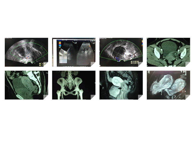
CASE 429: DOUBLE UTERUS, Dr PHAN THANH HẢI- Dr TRẦN NGÂN CHÂU, MEDIC MEDICAL CENTER, HCMC, VIETNAM.

$
0
0
Woman 48yo, PARA 2002 , detected herself one prolapsed mass from her vagina 1 year ago, no pain, no fever but with SUI [ stress urine incontinence] syndrome.

Ultrasound of pelvis by the transcutaneous: (US1) uterus normal size,


by via TVS ( US2, US 3) detected one mass at lateral left uterus, hypoechoic, size 7 cm look-liked second uterus.


CT scan of pelvis:  CT 1 : this mass at left site uterus, hypodense like fatty tissue.


CT2, CT3 the mass is anterior the urinary bladder.


 MRI 1, MRI 2 in sagittal section, this tumor is like a second uterus.


What is your suggestion?

CASE 430: FACIAL EDEMA, Dr PHAN THANH HAI, Dr LE NGOC VINH [MEDIC CA MAU], MEDIC MEDICAL CENTER, HCMC, VIETNAM

$
0
0


Woman 33yo, from Ca mau province, with history onset one year ago, fever and some red macula appeared at abdominal skin that biopsy result of macula was lipoma. But it is not in stop of progress, a lot of red macula were getting more over 2 legs and upper arms to her right face.
In palpation, red macula is hot and induration (Photo1,2.3).


Ultrasound scanning of  her right face represents edema of subcutaneous fatty layer, no hypervascular.


CT scan of the face shows normal bone and subcutaneous edema of her right hemifacial side.


MRI also detected facial edema of subcutaneous fatty tissue (MRI 1,2).


Blood tests= pancytopenia, 
negative ANA test and anti-dsdna, very high ferritin >2,000, LDH =2,581 UI,  beta2 microglobulin=  3,701,  CRP = 64 ng/mL, c3 =122 (normal), c4=  66 (n: 15-450), kappa and lambda  not detectable.

What is your suggestion for diagnosis of this hemifacial edema case?


CASE 431: BIG GASTRIC TUMOR, Dr PHAN THANH HẢI, MEDIC MEDICAL CENTER, HCMC, VIETNAM

$
0
0


Man 44 yo with one month complaining of epigastric distention but normal gastroendoscopy.  
Ultrasound  detected  big epigastric mass,  looked like  left hepatic tumor with size of 15 cm,  central necrosis (US 1, US 2).


MSCT CE represented that this tumors is  nearby  left lobe of liver  and  deplaces  gastric fundus [CT1, CT 2,  CT 3] with central  necrosis.


Blood tests with all cancer markers are normal. Wako test is  negative.  
Operation  removed  this big tumor   which came from  gastric wall.


Microscopic is GIST with high malignant potential.  


CASE 432: A K I by STONE, Dr PHAN THANH HẢI, MEDIC MEDICAL CENTER, HCMC, VIETNAM

$
0
0


Man  51 yo with acute  pain at  right flank, type colicky  pain. History of being treated  renal stone of  left kidney by operation and ESWL for 2 years.
Emergency  ultrasound  detected right and left kidney hydronephrosis (US 1, US 2).


CT scanning  with CE:  CT 1= kidneys no CE.


CT 2 with CE,  arterial phase.


CT 3,  venous phase.


CT 4,  delay phase.


CT 5,  frontal view.


 CT 7,  3D view.


Blood test: EGFR=  23mL/s.

Discussion: Ultrasound  scanning in acute renal colic crisis cannot make diagnosis of  A K I ( acute kidney insufficiency);   CT  non CE  with HU  low and CE phase in delay  secretion that suspected AKI.

 Emergent  operation was done  for removing  of the stuck stone in right ureter for this case.


CASE 433: GEANT RETROPERITONEUM LIPOMA, Dr PHAN THANH HẢI, MEDIC MEDICAL CENTER, HCMC, VIETNAM.

$
0
0


Woman 44yo nullipareous, with abdomen  distention, and  clinical  suspected   ovary tumor.
US scanning of abdomen detected  all bowell loops in deplacement to left flank.

US 1: epigastric scanning detected  right kidney near left lobe liver.


US 2: big solid mass, hyperechoic  like  fatty tissue.


US 3, US 4  color Doppler  of this mass showed from  retroperitoneum which extended from the epigastric  to pelvis but  no deplacement of abdominal aorta.


MRI  with  gado:  This  big mass is in retroperitoneum deplaces right kidney to liver (MRI 1, MRI 2, MRI 3,  MRI 4 , MRI 5).  


Radiologist reports   that is retroperitoneal lipoma or retroperitoneal liposarcoma.
Operation removed this mass easily.(see macro).


Wait for microscopic report.

CASE 434: TESTIS TUMOR, Dr PHAN THANH HẢI - Dr LÊ TÀI, MEDIC MEDICAL CENTER, HCMC, VIETNAM.

$
0
0

Man  36yo with right testis tense.
Ultrasound scanning of  right scrotum  detected big testis focal lesion,  round,  size of 3cm
US 1  color doppler  not  hypervascular, well bordered.

US 2 CDI: hypovascular tumor.

US 3  elastoscan of  this tumor is inhomogeneous with some parts very hard.


MRI  with gado: MRI 1, 2, 3: this tumor  very low gado enhanced.


Radiologist suggested epidermoid  cyst.
Blood tests =  normal   AFP  and   HCG.
Operation   resection of  right testis (see  macro 1, 2).


Microscopic report is epidermoid cyst.


CASE 435: CERVICAL LYMPH NODES, Dr PHAN THANH HẢI, MEDIC MEDICAL CENTER, HCMC, VIETNAM

$
0
0

Female patient 23 yo detected  submandibilar  nodes with history of recurrent sore throat. And some palpable  left cervical  nodes along  of SCM. No fever, nor pain.
US 1: hypoechoic, no hilus left submandibular nodes.


US 2: scanning  along  left site SCM, small hypoechoic nodes.


US 3: hypervascular  color Doppler .


US 4. high flow vascular  intranodes without defected area.


US 5: spectral Doppler  high RI and PI.


US 6: elastoscanning of  inhomogeneous nodes, central low kPa.


Biopsy of  the big submandibular node.
Blood tests=  normalWBC, CRP,  but high beta microglobuline 3200 UI, and  normal LDH, ferritine.
What do you suggest  for the case?


CASE 436: SOLITARY KIDNEY, Dr PHAN THANH HẢI, MEDIC MEDICAL CENTER, HCMC, VIETNAM

$
0
0

Man 32yo  with acute pain at  right renal fossa.
Emergency ultrasound:
US 1 = right kidney  hydronephrosis, no left kidney.
US 2 =  crossed section of  right kidney.
US 3 =  right  ureter dilated and stone of 1.5 cm in diameter.
US 4 = color Doppler  twinkling artifact  with stone.


US 5 = in  urinary bladder , urine jet only on right site.


US 6 =  cystic mass at the left wall of urinary bladder.


MSCT with CE=  
CT1:  fron tal view,  right  kidney  hydronephrosis no left kidney.
CT 2:  sagital view   the stone in right ureter.

CT 3:  3D view.
CT 4:  vascular malformation of agenesis of left kidney.


CASE 437: LIVER TUMOR, Dr PHAN THANH HẢI, MEDIC MEDICAL CENTER, HCMC, VIETNAM

$
0
0

Female patient 16 yo with epigatric pain.

Blood test=  HP positive.
Ultrasound  detected  left liver tumor.

US 1 =  longitudinal scanning of  left lobe of liver:   solid tumor , size  of 10 cm with central necrosis.

US 2 =  subcostal scanning :   tumor covers left liver lobe.

US 3  = color Doppler :  vascular supplying  of  this tumor.


US 4 =  elastoscanning of this tumor = 10,5-15 kPa.  


MSCT with  CE: CT 1, CT 2 =  artery and vein phases, CT 3 =  frontal view of this tumor from left liver.


Blood tests=  no infested HBV, HCV, Wako tests 3 negative.


What is your  suggestion for diagnosing of the case ?

CASE 438: RENAL HILUM TUMOR, Dr PHAN THANH HẢI, MEDIC MEDICAL CENTER, HCMC, VIETNAM

$
0
0

Female patient 21 yo detected high blood pressure of 17/10 cmHg.
Ultrasound of abdomen detected  one mass at  upper pole of left kidney with  size of 3.5 cm. This mass  covers  left border of aorta, left renal hilum and adrenal fossa (US 1, US 2, US 3).


MSCT with CE= CT 1, CT 2,  CT 3, CT 4 (3D vascular)=    this tumor  covers the hilar kidney, very high CE enhancement. Radiologist says  adrenal tumor.


Blood test=catecholamine blood and  24 hrs urine analysis detected not abnormal
Metanephrine  blood =102 unit  (n= 90),  in urine = low 42 unit.
Operation  by laparotomy=

 Picture 1= this tumor  covers  the  left renal hilum.


Picture 2= Nephrectomy with tumor specimen.


CASE 439: URINARY BLADDER TUMOR, Dr PHAN THANH HẢI, MEDIC MEDICAL CENTER, HCMC,VIETNAM

$
0
0

Man 69 yo with  hematuria. Ultrasound  detected 2 tumors in urinary bladder (UB), one in fundus of UB, other one in prostate  ( US 1,  US 2 ), US 3=  color Doppler with twinkling artirfact due to calcification, US 4 = cystic tumor in left kidney.


MSCT with CE of  urinary tract confirmed left kidney cystic tumor and urinary bladder tumor.


By via cystocopy biopsy result is transitional cell carcinoma of UB.


Viewing all 624 articles
Browse latest View live