Quantcast
Channel: VIETNAMESE MEDIC ULTRASOUND
Viewing all 626 articles
Browse latest View live

CASE 681: PARATHYROID TUMOR, Dr PHAN THANH HAI, Dr JASMINE THANH XUAN, Dr HO CHI TRUNG, Dr LE VAN TAI, Dr LE TUAN KHUE, Dr TRAN THANH CUONG, MEDIC MEDICAL CENTER, HCMC, VIETNAM.

$
0
0

A 33 year-old male patient with dysuria 

His history noted 6 times of long bone fracture, and renal stone and double JJ sonde on the right kidney.

Ultrasound detects some renal stones on 2 sides. And with herself experience sonologist reveales a left parathyroid tumor next to left lobe of thyroid.


Lab data and X-Rays show a case parathyroid tumor with bone complications.


Osteoporosis in severe level.


Surgery removes the parathyroid tumor  and the PTH goes down after that.


So the patient suffers from the renal stones, the seveve bone complications from the first time of bone fracture due to loss of information of the cause of his bone fracture.


CASE 682: RIB CHONDROSARCOMA, Dr PHAN THANH HAI, Dr HO CHI TRUNG, MEDIC MEDICAL CENTER, HCMC, VIETNAM.

$
0
0


A 56 male patient with right shoulder pain for a half of month.

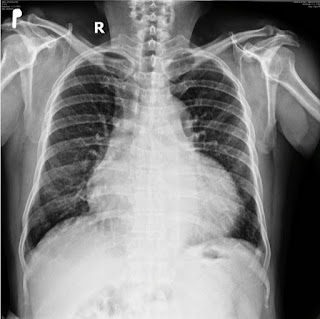
Chest X-Ray and ultrasound show a #3.1x2.5 cm right posterior legion of 7th rib  with hypoechoic pattern, neovascular signals and moderate of hard code on elastography ultrasound.


CT confirms and  suspects malignancy to the # 3.5 cm diameter of rib lesion with erosion of upper border of the 7 th rib which is strongly capturing CE.


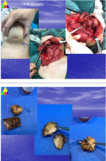
Surgery removes the tumor and the histopathologic report is mesenchymal chondrosarcoma.


It is a rare entity of cancer of cartilage in middle aged patient  low metastase. 

CASE 683: A TB CASE: Dr PHAN THANH HẢI, Dr TRẦN THỊ TRÚC PHƯƠNG, MEDIC MEDIC CENTER, HCMC, VIỆT NAM

$
0
0

 A 29 year-old female  patient in a general chek-up for a preparation of a study abroad.

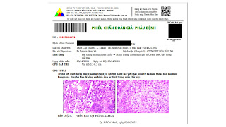
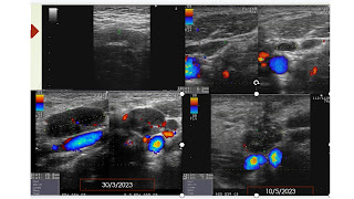
Ultrasound detects lymph nodes in her mesentery in the abdomen and on her neck # 5-25 mm in suspecting TB infiltrating type. Also a slight thickening of wall of colon is noted.


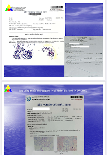
Report of the neck lymph node full biopsy is an TB  infected node.


A colonoendoscopy performs shows findings of ulcerative colitis from the cecum to the transverse colon due to TB infected.


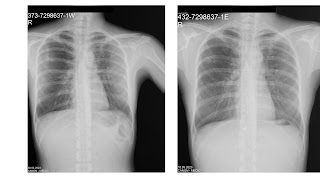
A TB regimen is planned for the patient, and during 2 months of TB management, her status is getting better, that is proven by the clear chest X-Ray and decreasing of lymph node in size and numeration.


CASE 684: PRIMARY BREAST SARCOMA, Dr PHAN THANH HẢI, Dr LÊ THÔNG LƯU, MEDIC MEDICAL CENTER, HCMC, VIETNAM

$
0
0

A 61 year-old female patient suffers from 2 tumors of her right breast which are abandoned for 2 years by her husband's deadly illness.

Ultrasound  and elastography technique notes 2 BI-RADS 4B tumors of her right breast.


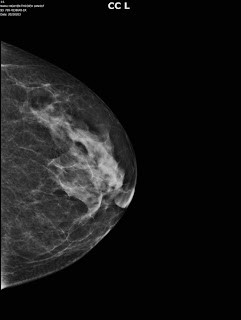
Mammography notes a non symetric sign in superiolateral region of the right breast.


MRI confirms the 2 BI-RADS 5 right malignant breast tumors: # 34x28 mm and # 21x 27 mm, spiculated border, high signals on T2W2 and medium signals on T1W1, captured contrast media type 3-2.


But the report of histoimmunology is a breast sarcoma while axillary lymph nodes are not in malignancy.


Surgery is done in large field, no mastectomy nor lymph node curetage due to the sarcoma tumor characters.


As no clue of gene mutation, the patient is planned for 3 months of 54Gy dosages in 27 times of  radiation therapy.

DISCUSSIONS:

Breast sarcome is a rare mesenchymal breast tumor (<1% cancer breast tumor). MRI,  mammography and ultrasound could not differentiaze breast sarcoma from other breast cancer tumors.
Core biopsy and histoimmunologic exam are keys of diagnosis.
Surgery could save patient life that sarcoma invades in situ and rarely via the blood stream. Chemotherapy and radiation may be managed in case of metastase and spreading. Liver, lung, bone marrow and recurrent breast tumor may happen in the first 2 years.  The 5-year survival rate reported in the literature ranges from50% to 64% for the breast sarcoma.


REFERENCES:


CASE 685: ANEURYSM of LEFT ILIAC VEIN and CORKSCREW of LEFT EXTERNAL ILIAC ARTERY, Dr PHAN THANH HẢI, Dr NGUYỄN ĐỨC DUY LINH, Dr NGUYỄN NGỌC XUÂN GIANG, BSc TRƯƠNG TẤN PHÁT, MEDIC MEDICAL CENTER and BINH AN HOSPITAL, KIEN GIANG PROVINCE, VIETNAM.

$
0
0

 A 58 year-old male patient in general check-up is revealed incidentaĺly by ultrasound an aneurysm of left iliac vein  # 48x66 mm without thrombosis. In addition, the left external iliac artery dilates #15-21mm appears in tortuosity like a corkscrew.


MSCT 64 confirmes the findings of the aneurysm of left iliac vein  # 48x66 mm without thrombosis. 


And in 3 D reconstruction the corkscrew of left external iliac artery appears clearly in mild dilatation without damage of its wall beside the aneurysm of the left iliac vein.

The corkscrew of external iliac artery is an anatomic variant incidentally revealed by CTA but a skilled sonologist could detects it with experience oneself. However, the key of this case is the aneurysm of iliac vein which leads to find out an uncomplicated aneurysm and the arterial tortuosity of the external iliac artery close by. But the cause of the left iliac vein aneurysm is unknown.

Aneurysm of iliac vein is a rare entity which appears in men and on the left side. And in contrast, female gender is a predisposing factor of the arterial tortuosity.


The patient is planned to the conservative management.

REFERENCES


:


CASE 686: GASTRODUODENAL ARTERIAL ANEURYSM, Dr PHAN THANH HẢI, Dr PHAN THỊ HƯỜNG, Dr HỒ KHÁNH ĐỨC, MEDIC MEDICAL CENTER, HCMC, VIETNAM.

$
0
0


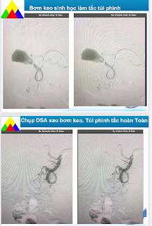
A 64 year-old HTA diabetic male patient is detected an arterial aneurysm # 35x34 mm by ultrasound maybe a gastroduodenal arterial aneurysm.


MSCT later confirms the 35x32 mm arterial aneurysm of gastroduodenal artery.


The patient goes through an endovascular aneurysm repair (EVAR) by biological glue.
The patient remains well post-op.

But ultrasound could note the vestige of aneurysm # 34x31 mm without any Doppler signal and flow after 8 months.

CASE 687: SUBMANDIBULAR TUMOR, Dr PHAN THANH HẢI, Dr TRẦN LÃM, MEDIC MEDICAL CENTER, HCMC, VIETNAM

$
0
0


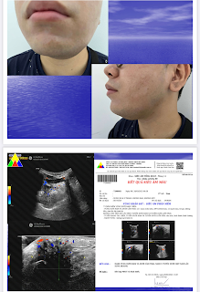
 A 27 year-old male patient suffers from left submandibular gland for 3 years.

Ultrasound notes a left submadibular gland tumor.


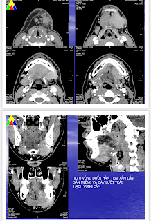
MSCT confirms the left submandibular  that invaded the mouth floor and node.


But result of FNAC has only an amount epithelium cells and RBC.  And in the second time maybe a pleomorphic tumor.

Goes through a surgery, the specimen is a malignant submandibular gland tumor, mucoepidermoid carcinoma low-grade.


CASE 688: ASYMMETRICAL HYPERTROPHY of the HEART, Dr PHAN THANH HẢI, Dr NGUYỄN DINH, Dr ĐÀO XUÂN DUNG,MEDIC MEDICAL CENTER, HCMC VIETNAM.

$
0
0


A 59 year-old arterial hypertension male patient with a chest pain in taking alcohol from October 2019. He came Heart Institut HCMC with a note of infarctus of myocardium from Cantho province.  


But MSCT in Hoan my hospital Cantho province proves a slight stenosis 30% of coronary arteries. 

In COVID 19 pandemic, he goes around the private clinics with his HTA status and his heart but nothing change.

In May to July 2022 he goes to Medic Can tho and then Medic Hoa Hao Center in HCMC.

An coronary CTA of this second time shows slight stenosis under 30% and slight pericardial effusion but notes a hypertrophic cardiomyopathy of his heart.


At last Cardiac MRI confirms an asymmetrical hypertrophy of the heart.


He gets well with the management of  hypertrophic cardiomyopathy.


REFERENCE


CASE 689: RECTUM WALL GIST, Dr PHAN THANH HẢI, Dr LÊ TUẤN KHUÊ, MEDIC MEDICAL CENTER, HCMC, VIETNAM

$
0
0

A 56 year-old female patient with 6 month constipation and bleeding from her anus  for 2 weeks.

Rectal endoscopic result is a hemorrhagic rectal tumor which is far from anal orifice 20 millimeters.

MRI confirms a 53x41 mm tumor of the rectal canal.


A transperineal biopsy with ultrasound-based guiding is done for a 42x48x44mm vascularised solid mixed tumor.


Result of histoimmunology is a GIST tumor of  rectum wall.


The female patient is planned for a surgery and chemotherapy.

CASE 690: APPENDICULAR ABSCESS, Dr PHAN THANH HAI, Dr NGUYEN NGOC XUÂN GIANG, MEDIC MEDICAL CENTER - BINH AN HOSPITAL KIÊN GIANG, VIETNAM

$
0
0

A 56 year-old female patient with umbilical pain for one week, comes to Binh an hospital after 4 days in constipation.

Ultrasound detects an abscess in RLQ but could not rule out a PID. Although sonologist notes an appendicular abscess but could not reveal the inflammed appendix into the pelvic abscess.


MSCT confirms a 60x80 mm  right pelvic abscess contrast captured and edema of the wall of terminal jejunum. The report is pelvic abscess maybe an appendicular abscess.


Endoscopic surgery drained the pelvic abscess and the patient remains well.


CASE 691: LEFT ADRENAL TUMOR, Dr PHAN THANH HẢI, Dr VÕ THỊ PHƯƠNG TRINH, MEDIC MEDICAL CENTER, HCMC, VIETNAM

$
0
0


A 48 year-old female patient without arterial hypertension signs is incidentally detected a tumor of adrenal gland #20×22 mm by ultrasound.


MSCT confirmes  a 16x22 mm left adrenal benign tumor  which HU before CE is 69 and HU post CE =26. CE discharge is over 50% that may proove the benignity of the tumor.


4 months after the left adrenal tumor is removed by endoscopic surgery.


Result of pathology is a benign adrenal tumor with thin fibrous capsule.


CASE 692: Five Year HCC post-op, Dr PHAN THANH HẢI, Dr DƯƠNG NGỌC THÀNH, MEDIC MEDICAL CENTER, HCMC , VIETNAM.

$
0
0

 A 71 year-old female patient with her history of a liver tumor which was quite difficult diagnosed 5 years before.

She had been go through ultrasound, MSCT  and liver biopsy 2 times in 2 other hospitals, but the results were benign hepatic tumor and no clue of infected HCV and HBV.

In Medic Center, ultrasound noted a 50 milimeter right liver hemangioma and MRI  with gado was an AML but not ruling out HCC.


But HCC risk tests with WAKO test were positive and positive antiHBe in that time.


After 2 months of the five year before to examine the liver tumor, the HCC  diagnosis was made and the tumor was removed surgically when the patient aged 66.


Histopathological report was a poor differentiazed carcinoma of liver with fatty degeneration  and hepatic chronic inflammation.

And 5 years went by the female patient remains well. The right lobe of liver exists a fibrosis scar.


The prognosis of HCC patient will be well if an effective management performs successfully in time for the female patient [Acceptable 5-year survival].

CASE 693: ADVANCED MALIGNANT MESENCHYMAL CELLS of PROSTATIC TUMOR, Dr PHAN THANH HAI, Dr NGUYEN MINH THIEN, MEDIC MEDICAL CENTER. HCMC, VIETNAM.

$
0
0

 A 66 year-old male patient with necrotic periaortic lymph nodes and microscopic blood urine on abdominal ultrasound comes to Medic for a prostate biopsy. PSA value is 1.38mg/mL.

By via TRUS it exists a #55x43x62 mm big prostate, loss its capsule and distorsion of prostatic structure. The hole prostate is noted many hard sites on shear wave elastoultrasound.


12 specimen prostate biopsy performed and 16 items histoimmunopathologic report is a non specialized malignant mesenchymal cell of prostate tumor (PCa) on a chronic TB old inflammed based structure.


This is a rare PCa malignant mesenchymal cell  of prostate tumor may happen on 1/1000 cases.


CASE 694: SPONTANEOUS SPLEEN LACERATION, Dr PHAN THANH HẢI, Dr NGUYỄN NGHIỆP VĂN, MEDIC MEDICAL CENTER, HCMC, VIETNAM.

$
0
0

A male patient 56 year-old  with one day of pain at left side of abdomen, obesity, and five-year chronic leukemia.  His history is noted splenohepatomegaly and cirrhosis and controlled type 2 diabetes. 

At Medic Center ultrasound detects free fluid in abdomen and big spleen with hematoma inside which makes thought about a spontaneous spleen laceration without any injured causes in spite of spleen infarction.

MSCT  with CE confirmes a spleen rupture with subcapsular hematoma and free fluid in the abdomen.


Surgery in emergency is done to remove the # 180x120x80 mm spleen and hemostasis procedure.
At the middle of anteromedial face of spleen it exists a #50 mm long  and 30 mm deep laceration line. Bloody fluid in abdominal cavity is an amount of 1.8 liter.
The patient goes through successfully the operation  but comes back with an 80x89 mm spleen bed abscess which is solved by medical management.


Chronic leukemia and obesity and DM maybe the causes of a spontaneous spleen laceration.

CASE 695: Fasciola hepatica, UNUSUAL CAUSE of THICKENING of G I TRACT, Dr PHAN THANH HẢI, Dr TRẦN NGÂN CHÂU, Dr LÊ ĐÌNH VĨNH PHÚC, MEDIC MEDICAL CENTER, HCMC, VIETNAM.

$
0
0


Two cases of infected Fasciola hepatica whose larva migrants having in the same time some hepatic lesions and thickening of duodenum (2023) and right colon wall (2017) that are noted at Medic Center. 

CASE ONE:  A male patient 45 year-old with history of thyroid cancer in 2013 goes to a hospital as nausea, abdominal pain without fever after a ceremony buffet.  Ultrasound detects hepatic lesions and MRI later reveals lesions in caudate lobe of liver and duodenum D3 wall thickening that is thought a case of infiltration of lymphoma on GI tract and liver.

But lab data notes raised eosinophil (48%) and positive Elisa tests for Fasciola and Gnathostoma.
Ultrasound of Medic Center confirmes liver lesions  and  D3 wall thickening  that maybe concludes due to infected parasites.


After 6 weeks managed by medical parasite drugs for Fasciola  the male patient remains well; liver lesions reduces size and duodenum wall gets normal on ultrasound and abdominal MSCT and getting downed eosinophil proportion.


CASE TWO: 

A female patient with Fasciolasis lesions in her liver and her right colon wall thickening in the same time which were detected by ultrasound and MSCT. 


Endoscopic biopsy of colon result was epithelial inflammation with eosinophil white blood cells.


She was managed successfully as Fasciola visceral larva migrants.
Larva migrants, especially for Fasciola sp, have a classic site in liver and biliary tree in acute phase and chronic phase. 
It exists two cases of larva migrants going outside the liver and biliary tree, to GI tract. If having lesions of it in liver or biliary tree one should not forget to find out larva migrants in another place.

REFERENCE:

Ultrasound and Fascioliasis at MEDIC CENTER, Vietnam (slideshare.net) Oct, 21, 2013


CASE 696: PRIMARY CLEAR CELL HCC, Dr PHAN THANH HẢI, Dr NGÔ THỊ HUYỀN TRANG, MEDIC MEDICAL CENTER, HCMC, VIETNAM

$
0
0


A male patient 53 year-old with tumor in right lobe of liver and negative WAKO tests.

Ultrasound notes a 55x42 mm liver tumor in segment 7, nearly homogeneous, well-limited,  poor vascularised and elastographic ultrasound SWE harder 5-fold than hepatic parenchyma: 29 kPa in comparison to 6.3 kPa.


MRI with Primovist confirms a 50 milimeter clear cell HCC (CCHCC). T2 CE captured signals are higher than liver parenchyma and  lower than on T1. 


Biopsy results of tumor is an HCC well differentiazed.


REFERENCE:


Hepatocellular carcinoma (HCC) is a common cancer world-wide with a higher incidence in Asia. Clear cell variant of HCC (CCHCC) has a frequency ranging from 0.4% to 37%. The presence of 90-100% clear cells is rare.


CASE 697: STOMACH GIST, Dr PHAN THANH HẢI, Dr PHAN THỊ HƯỜNG, MEDIC MEDICAL CENTER, HCMC, VIETNAM.

$
0
0

 A 47 year-old female patient with a gastric tumor at lesser curvature which is spontaneously revealed by ultrasound.

But gastroendoscopy result is only a gastritis.


Later MSCT confirms  a 50x58 mm gastric wall tumor maybe a stomach GIST. The tumor captures CE inhomogeneously and goes out in compression organs around in abdominal cavity.


Surgery removes the gastric tumor and the histopathological result is gastric GIST.


REFERENCE:



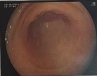


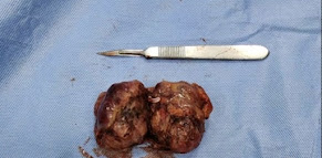


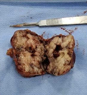


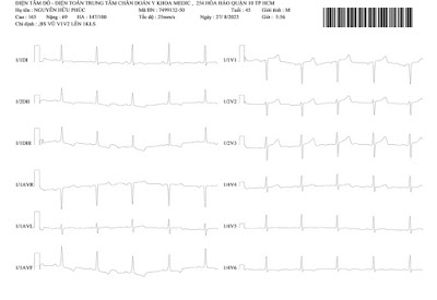


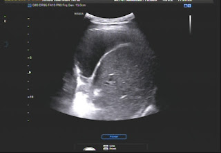
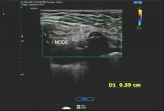

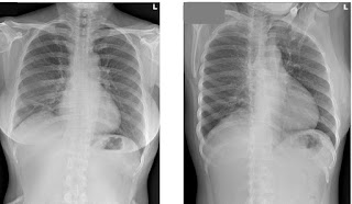


A 53 yearale patient with a gastric tumor which is spontaneously evealed 


CASE 698: HTA YOUNG PATIENT with 2 RENAL ARTERIES each side, Dr PHAN THANH HẢI, Dr PHAN THANH HẢI PHƯỢNG, Dr HỒ KHÁNH ĐỨC, MEDIC MEDICAL CENTER, HCMC, VIETNAM

$
0
0


Chest X-RAY film is normal and so his EKG is ischemic heart disease.


Lab data is normal.


At last, DSA  detects 2 renal arteries each side that belongs to a renal artery malformation.


REFERENCE


Renal arteries are a pair of lateral branches from abdominal aorta. Normally each kidney receives one renal artery. However, accessory renal arteries can also exist. The normal renal arteries enter the kidney through its hilum where as the accessory renal arteries might enter the renal artery through the hilum or through the surfaces of the kidney. Knowledge of the variations in the renal arteries is important for urologists, radiologists and surgeons in general.


Accessory renal arteries are common in 20–30% of individuals, usually arising from the aorta above or below the main renal artery. The variation in the number of arteries is because of persistence of lateral splanchnic arteries or due to the persistence of blood supply from lower level than normal.


CASE 699: DIFFUSE LARGE B CELL LYMPHOMA and WALDEYER'S RING, Dr PHAN THANH HẢI, Dr DƯƠNG XUÂN TÙNG , MEDIC MEDICAL CENTER, HCMC, VIETNAM

$
0
0

A male patient 41 year-old with multiple cervical nodes and sore throat as tonsillitis.


Chest X-RAY notes left lung lesion.


Ultrasound detects many lymph nodes  # 17-15-13 mm without nodal hilus, solid, hypoechoic on his neck and in the abdomen : at liver hilus :27mm, mesenteric,  pelvic: 17-21 mm and a splenomegaly :141mm. Results of ultrasound notes a multiple lymph nodes in cervical, supraclavicular and abdominal region that leads to a diffuse lymphoma. 


Biopsies of tonsils and pharyngeal cavum results are lymphoma infiltration without immunohistochemical staining.


ENT examinations results are many lesions of tonsils and oral cavity and Waldayer's ring.


Biopsy of right tonsil ulcer for ruling out cancer and immuohistochemical staining result is diffuse lymphoma type large B cell.


Patient goes through a chemotherapy planning for lymphoma. Cervical nodes reduce their sizes with effective management. 


REFERENCES:


CASE 700: RIGHT THORACIC WALL TB ABSCESS, Dr PHAN THANH HẢI, Dr JASMINE THANH XUÂN, Dr HỒ CHÍ TRUNG, Dr LÊ THÔNG LƯU, MEDIC MEDICAL CENTER, HCMC, VIETNAM.

$
0
0

 A 45 year-old  female patient with right thoracic painful swollen area for 5 months.


Ultrasound detects right pleural effusion, thoracic wall mass which contains rib cartilage destruction, close by pleural wall thickening at 4 th intercostal space, and local lymph nodes.


Chest X-RAY  shows right pleural effusion and nothing about thoracic wall. 


MSCT  confirms a right thoracic wall lesion and right lung NAD.


FNAC and core biopsy of right thoracic wall results  think  about TB inflammed lesion with ADA raises slightly in right pleural fluid.


So it exists a painful right thoracic wall for 5 months and evidents belongs to a TB infection without primary lung lesion.

It will be planned for a TB regimen in TB and Lung hospital.

Viewing all 626 articles
Browse latest View live