Quantcast
Channel: VIETNAMESE MEDIC ULTRASOUND
Viewing all 626 articles
Browse latest View live

CASE 599: TESTICULAR CANCER MIMICKING TESTIS TORSION , Dr PHAN THANH HẢI, Dr NGUYỄN MINH THIỀN, Dr MAI BÁ TIẾN DŨNG, MEDIC MEDICAL CENTER, HCMC VIETNAM

$
0
0

Male patient 31 yo, with sudden pain at left scrotum for 2 months had been treated as epidydimitis but treatment failed. He came to Medic for reexamination because swollen scrotum and testicular pain.


Ultrasound at Medic Center detected swollen left testis  with edema of epidydimis and hypervacularization. Testicular axis turned horizontally and left testis was inhomogenous with cystic necrosis and no vascular signal mimicking a left testicular torsion.


MRI of left testis #  60x85mm, inhomogenous signals that existed fluid and blood inside but captured a few of contrast. Edema of epidydimis and spermatic cord. No spermatocele.

Lab results showed no sign of inflammation, beta HCG, AFP, LDH raising that lead to think about a testicular  tumor non seminoma.


Operation removed left testis. It looks like tumor on macroscopic view. Histopathologic result is testicular embryonic carcinoma.


Post surgery one day,  blood tests dropped=  AFP, Beta HCG and LDH   ( AFP= 62, beta HCG= 8.9, LDH= 419). Normal. Chest XRray .  


DISCUSSION= Diagnosis of left testicular tumor based on patient history, age, beta HCG, LDH and AFP raising. No hypervascularizing of left testicular tumor maybe due to thrombosis of vessels in spermatic cord that could make mistake for ultrasound and MRI.


CASE 600: MIRIZZI SYNDROME, Dr PHAN THANH HẢI, Dr PHAN NGUYỄN THIỆN CHÂU, Dr NGUYỄN NGHIỆP VĂN, MEDIC MEDICAL CENTER, HCMC, VIETNAM

$
0
0

 Male patient 62yo with RUQ pain for 1 month failed  managed as gastritis. Ultrasound at local hospital detected GB stone so the patient came to MEDIC to reexamination.


Ultrasound at Medic Center detected big GB stone # 24mm and CHB dilatation, thought about GB stone and Mirizzi syndrome.


MSCT confirmed later GB stone in cystic duct and Mirizzi syndrome.


Lab tests CA 19-9=145,5U/mL, Bilirubin raising esp direct bilirubin.


Patient was sent to Binh dan hospital to manage the GB stone.

Bình dan MRI confirmed GB stone and Mirizzi syndrome.


Surgery was done to remove GB stone, cholecystectomy and Kerh drainage for C H D.


CASE 601= MALIGNANT LIVER TUMOR BUT WAKO TEST NEGATIVE, Dr PHAN THANH HẢI, Dr DƯƠNG NGỌC THÀNH, MEDIC MEDICAL CENTER, HCMC, VIETNAM

$
0
0


Male patient 43yo with HBV infection for years but only follow-up now detected right lobe indeterminated tumor #30x23mm at subsegment VIII on ultrasound. Doctors thought benign tumor because of WAKO test negative.


MSCT confirmed a focal fatty infiltration on right lobe in different diagnostic of a liver tumor.


One month later, MRI with Primovist detected  liver tumor at VIII segment, T1 low  tumor signal than mesenchymal signal, but T2 higher than liver. Wash-out is typically same HCC  (7sec, 7min and 30min).


The tumor is close to IVC and between right and middle  hepatic veins that will invade vessels if delayed management.


Open surgery to remove tumor in anterior lobe of liver. Histopathologic report is liver cell carcinoma, trabeculated pattern.


CONCLUSION: MRI with Primovist is best choice for small liver HCC with negative WAKO test.

CASE 602: HEPATIC ABSCESS MIMICKING HCC in CHRONIC HBV PATIENT, Dr PHAN THANH HẢI, Dr TRẦN THÙY TRANG, MEDIC MEDICAL CENTER, HCMC, VIETNAM

$
0
0


Male patient 36yo with HBV infection  known by himself since November 2019 without treatment.

Ultrasound at Medic Center showed a 39x22mm mass in right lobe on chronic hepatitis # F 3.

      US 1: Ultrasound detect an ill-defined, hypoechoic mass #4cm (with a small cyst in center) in hepatic segment VI.


      US 2: Superb microvascular imaging (SMI) shows small blood vessels in tumor.


      US 3, 4: Elastography shows that the stiffness of the tumor and its outline are harder than the liver parenchyma.


Lab results: HBsAg +; HBeAg +; HBV DNA =62,953U/mL; AFP=5.97. Wako tests=AFP L3 <0.5, PIVKA II(DCP)=19


MRI with Gado thought about 36x30mm HCC= T2 higher signal than liver; T1 lower signal. Gado caught more in arterial phase, in late phase lower signal than liver.


Operation removed the liver mass 


and histopathologic report said liver tissue inflamed with majority of eosinophil leucocytes.


CASE 603: DVT in ANEMIA PATIENT with ILEUM TUMOR, Dr PHAN THANH HẢI, Dr NGUYỄN NGHIỆP VĂN, Dr TRẦN THỊ THANH NGA, MEDIC MEDICAL CENTER, HCMC,VIETNAM.

$
0
0

Male patient,55yo from Kien giang provincewith history of HCV infectiondiabetes and  

arterial hypertension suffered from swollen right leg which is due to DVT and stenosis of tibial artery of right lower limb on anemia status

He has been managed his anemia and venous thrombosis in a local hospital by folic acid and Rivaroxaban 10mg. 

In general examination, slight anemia. P=90b/min,TA=130/80mmHg. 


Lab results shows anemia status Hb=9.8g/dLHct=31.3%, dDimer=511.  

[maybe with a relation of cancer #21%]  


Ultrasound at Medic detected DVT right leg 

and ileum wall thickening #23mm that  MSCT confirmed later. 


MSCT=


Surgery removed bowel tumor and histopathologic report 

is medium differentiated invasive carcinoma. 


CONCLUSIONS=


In old patient with anemia and venous thrombosis should be find out GI tract tumor. 


CASE 604: E M S of GALLBLADDER NECK STONE, Dr PHAN THANH HẢI, Dr LÊ THANH LIÊM, MEDIC MEDICAL CENTER, HCMC,VIETNAM

$
0
0

Male patient 50yo with pain at right costal border in coughing.

Emergency medical sonography (EMS) detects small stone # 6mm at the GB neck and some cholesterol polyps. GB size = 76x37mm, slight edema of  GB wall with hypervascularization due to inflamation. Common bile duct is not dilated.


MSCT non CE confirmed stone at the GB neck and GB wall thickening.


Laparocholecystectomy removed gallbladder with many stones.


CONCLUSION= Emergency medical sonography is an  efficient tool for diagnosis  of a RUQ pain case but that is non radiation in comparison to MSCT.


CASE 605: SPLENIC ARTERY ANEURYSM, Dr PHAN THANH HẢI, Dr VÕ NGUYỄN THÀNH NHÂN, Dr HỒ KHÁNH ĐỨC, MEDIC MEDICAL CENTER HCMC, VIETNAM

$
0
0

Female patient, 50yo, in general check-up by ultrasound incidentally had been detected a mass 23.5x25.55mm nearby the hilus of her spleen. This cystic structure has Doppler signal inside with zinzang sign. Sonologist thought about a cystic form of  aneurysm of splenic artery.


MSCT at Medic confirmed a sacciform aneurysm of splenic artery with calcifications,


Operation was done to remove the spleen and aneurysm.


Splenic artery aneurysm occurs in approximately 0.1% of all adults. It is estimated that 6% to 10% of splenic artery aneurysms will rupture, and 25% to 40% of those ruptures will occur during pregnancy, especially during the third trimester. Risk factors for rupture include portal hypertension and pregnancy.

Splanchnic Vessel Aneurysm at MEDIC
https://www.slideshare.net/hungnguyenthien/splanchnic-vesselaneurysms?qid=d9f6f654-e6c7-42ce-8104-5cd4b9f9a4bc&v=&b=&from_search=2


CASE 606: EXTRAHEPATIC MULTIPLE CHOLANGIOCARCINOMA, Dr VÕ HIẾU THÀNH, Dr NGUYỄN THỊ BÍCH NGỌC, Dr PHAN THANH HẢI, MEDIC MEDICAL CENTER, HCMC, VIETNAM.

$
0
0

Female patient 31yo, loss of weight, RUQ and epigastric pain for some months with unknown cause of dilatation of intra-extrahepatic biliary tree. 

At Medic Center, ultrasound shows a distention of gall bladder #132x62 mm without stone and some 10 - 17mm no vascular sign polyps. Furthermore there was a # 10 mm dilated common bile duct without stone downward to pancreas head

Intrahepatic biliary tree dilated slightly without stone. 


MRCP performed and detected CBD in dilatation then

collapsed near pancreatic headIntra and 

extrahepatic biliary tree dilated

Gallbladder wall irregular thickening with mass # 17mm. Nodes exist around pancreatic head.

Uncinate processus of pancreas head has high 

signal of contrast capture in T2FS. No dilatation of pancreatic duct. Radiologist thought about terminal part of CBD tumor invades uncinate processus of pancreas head. 


Pancreaticoduodenectomy operation of Whipple 

removed duodenopancreatic bloc. 


Histopathologic results are 1/ poor differentiated 

adenocarcinoma of CBD invades duodenum

pancreas and nerves and 

2/ poor differentiated adenocarcinoma of gallbladder  invades peritoneal epithelium and vesselsSomes metastatic lymph nodes 4/4 and 6/7 nodes. 


CONCLUSION= A rare case of multiple cholangiocarcinoma because it exists in young 31 yo female patient 

[usually in 50 yo or over patient]

 metastazing to pancreaslymph nodesnerves and vessels. Limits of technics of ultrasound makes ultrasound not reliable to find out cause of biliary duct obstruction and pancreas lesion than MRCP with contrastMaybe elastography ultrasound and endoscopic ultrasound could detect more but unfortunately that not in use in this interesting case. 


CASE 607: ISOLATED ABDOMINAL AORTA DISSECTION (IAAD) in YOUNG ALDULT, Dr LÊ THANH LIÊM, Dr PHAN THANH HẢI, MEDIC MEDICAL CENTER, HCMC,VIETNAM.

$
0
0

Male patient 38yo with sputum coughloss of weight #10kg, no fever, no abdominal painloss appetite for 2 months without trouble of passing out of water and waist

Gastroendoscopy shows gastritis and esophagitis

due to Candida. 


Normal chest-X-Ray. 


MSCT of chest and abdomen detected dissecting 

IAA from lower kidney part to left iliac artery while AA diameter suprarenal =21mm and infrarenal =22mm

Enlarged lymph nodes at both 2 lung hiliimediastinalleft axillary nodes

and in abdomen, lymph nodes of celiac artery, nearby 

pancreatic headand periaotic in epigastric area.

Thought about lymphoma infiltrating nodes. 


POC Ultrasound findingsSplenohepatomegalies 

and enlarged nodes of celiac arterynearby 

pancreatic headand periaotic in epigastric area. 


Thank to MSCT results, POCUS showed a dissecting isolated abdominal

aorta, d# 25 x 18mm from lower kidney to aortic bifurcation, which has  two lumens, right lumen with Doppler flow and no flow 

in left lumenRight and left iliac arteries with normal

lumen and Doppler flow. 


Video Clip= Doppler  isolated AA dissection.


Neck ultrasound  shows left side neck nodespoor  

echogeneicityloss of nodal hilus, no calcification 

nor necrosis sign

Nothing abnormal on thyroid scanning. 


Lab tests = Slight anemia,Hb=11.4g/dLLeucocytes= 8.95x10^9/L, normal FBG= 4.77 mmol/L, HP Test-IgM - IgG (Elisanegative; β2 Microglobulin = 4250 μg/L; HIV Elisa (+).


Biospy of neck nodes and histopathologic result 

Loss structure of nodeMany smallmedium and big size cellules in vessels with high endothelial cellshyalinized vessel walls

Many eosinophyl leucocytescytoplasms and 

epitheloid hystiocytes in the base.


Suspect T lymphoma on node specimen

Waiting for chemohistoimmunostaining. 


Conclusions 

1/ Pay attention of dissecting isolated abdominal aorta may exist in young patient.

2/ Complete examination and using all imaging

 diagnostic modalities may help detecting

 patient of risks and his/her illness. 


Reference:

Isolated Abdominal Aorta Dissection,

https://www.ncbi.nlm.nih.gov/pmc/articles/PMC3926414/

CASE 608: APPENDICOLITH, Dr NGUYỄN NGHIỆP VĂN, Dr VÕ NGUYỄN THÀNH NHÂN, Dr NGUYỄN PHÚ HỮU, Dr PHAN THANH HẢI, MEDIC MEDICAL CENTER, HCMC, VIETNAM

$
0
0

 Male patient 17yo, RLQ pain at 01:00 a.m. March 6, 2021.

Ultrasound at Medic Center at 10:00 a.m. detected an 33x10 mm swollen appendix at RLQ with fecolith =11 mm inside which has echo rich pattern and posterior shadowing. No free fluid around. A diagnosis of acute appendicitis with fecolith was made.


Lab results= WBC with neutrophil raised and CRP raised.


Later MSCT of abdomen confirmed an acute appendicitis with fecolith.


Patient was hospitalized  at 12:00 p.m. in Binh dan hospital. Endoscopic operation performed at 12:30 p.m..

And surgical macroscopic specimen (received via email at 03:00 p.m.).


Reference:

https://www.ncbi.nlm.nih.gov/pmc/articles/PMC3072212/


CASE 609: INTERESTING GASTRIC TUMOR, Dr LÊ THANH LIÊM, Dr PHAN THANH HẢI, MEDIC MEDICAL CENTER, HCMC, VIETNAM

$
0
0

Male patient 54 yo, with hemorrhagia due to gastric ulcer for 2 years. From late 2 months patient got pain  from neck to epigastric area, loss of appetite and weight. (Weight=58 kg, height=160 mm).

Ultrasound detected some nodes in both 2 lobes=10-38mm with halo sign and without Doppler signal. No thrombus in portal vein,  IVC and hepatic veins. None lymph node. Small amount fluid in pelvic area.


Stomach= Irregular thickening wall, d#27-69 mm of  nearly total gastric wall of corpus and fundus  that formed hypoechoic mass d# 161x166x163mm, hypervascular which takes wide place of lumen and  compressed cardia. Though about Gastric GIST with hepatic metastases.


MSCT with contrast confirmed  gastric GIST # 16 cm, adhesing around and liver metastases. Some low signal liver lesions 10-35 mm were in lower density than liver parenchyma. Corpus of stomach has lesion d= 16 cm with soft tissue density and has big ulcer at center. Lesion was adhesive and compressed around. Pelvic area has a little of fluid.


Blood tests= Severe anemia Hb 6.0 g/dl; Hct 25%; MCV 57.6 fL; MCH 13.9 pg; WBC slight raised=11.90 x 10^9/L; PLT high raised 794x10^9/L. HP Test-IgG (Elisa) POS 69.88 U/mL; HP Test-IgM (Elisa) POS 68.48 U/mL.


In Binh dan hospital, gastroendoscopy shows big  gastric tumor in fundus with deep ulcer having hard border and compresses lower 1/3 part of esophagus.


Histopathologic results= Infiltrating of lymphocytes and plasmocytes gastric mucosa proliferates fiber tissue and fibrosis. Masses of cells line in band with fusiform nucleii on base of fiber tissue. Follow up  GI GIST.

Waiting for histoimmunostaining.


CONCLUSION= Interesting diffuse appearance of entire gastric wall tumor helps diagnosing gastric GIST based on ultrasound and MSCT findings.

CASE 610: Multiple Brown Tumour. Dr Phan Thanh Hai Phuong, Dr Ly Van Phai, Dr Hoang Thi Thanh Phuong (Oncology hospital)

$
0
0

Patient came to clinic with history of 3 years worsen polyarthralgia. He has been diagnosed and managed as degenerative arthritis in BacLieu Province Hospital for a year. The pain localized in left shoulder, right knee, hips and left ankle. Patient also experienced general fatigue and weight loss (5kg a year). MSK Ultrasound is indicated as first tool, goes along with blood tests.

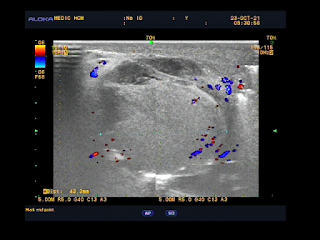
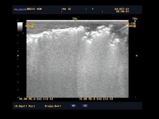
Ultrasound of right knee: shows patient area of pain doesn’t come from joint but the bone. It exists erosion of periosteal of proximal tibia and  hypervascularity in surrounding soft tissue. By switching to more penetrative linear probe, a hyperechoic bone mass is seen beneath periosteal erosion. Mass is homoechogeneity and has anechoic cysts inside.


Image 1: Erosion of periosteal of proximal tibia and hypervascularity in surrounding soft tissue


Image 2: A hyperechoic bone mass is seen beneath  periosteal erosion. Mass is homoechogeneity with  anechoic cysts inside. Second images shows normal articular cartilage and not fit for degenerative arthritis.

Ultrasound of left ankle:

Same condition as proximal tibia as in the distal end. Mass is better demonstrated with infiltrative border. During examination, patient feels imminent pain at place that probe compresses. Ankle joint is normal.


Image 3:  Mass is better demonstrated with infiltrative border in distal end of tibia

Ultrasound of shoulder:

Shoulder rotator cuff are normal. Area of pain is at left A-C joint. In comparison to normal right side, same apperance of bone mass revealed at clavicular end.


Image 4: Shoulder rotator cuff are normal. Area of pain is left A-C joint. In comparison to normal right side, same bone mass at clavicular end revealed.

Conclusion : Multiple distal bone masses suggest few differential diagnosis: Metastasis, Multiple myeloma metastasis.

Those could be pseudo-mass  coming from bone erosion in osteclastic hyperactivity  in hyperparathyroidism. Althought the lesions are common in middle diaphysis but not in the distal/proximal end. Radiologist did a quick check on the neck to rule out tumor of parathyroid gland.


Ultrasound of the neck: detected at right lower lobe of thyroid a parathyroid tumor, 3.7x1.4mm, enlarged with capsule and hypervascular on colour Doppler mode.


Image 5: Right parathyroid tumor, 3.7x1.4mm, enlarged with capsule and hypervascular signals on colour Doppler mode.


Image 6: Pelvis X-Ray: shows multiple scattered oval-shaped bone radiolucent, losing bone general density.


Image 7 : Right ankle XRay:  radiolucent oval-shaped lesion at distal tibia end in comparison to ultrasound.


Image 8:  Right ankle XRay:  radiolucent erosion lesion at distal clavicular end as compared to ultrasound.

Patient is preferred to endocrinologist and  full body scan to perform looking for bone fracture as common complication because patient suffering illness for a long time.


Patient blood tests confirmed diagnosis: hypercalcemia and normal RF quantitative. Serum PTH value elevated >1200


Patient underwent surgery to remove the tumor. Pathological result : Parathyroid adenoma.


After the surgery, PTH value drops to normal value, serum calcium also drops below normal line and had been got oral calcium supply as he discharged from hospital. 

Follow up on 3 months later, patient recovered and experienced no pain. He has already can go back to work.


Discussion and conclusion:

Primary hyperpaprathyroidism is caused by parathyroid tumor excretes PTH. That activates re-absortion calcium in kidneys, increase absorption in colons and bone loss. Osteoclastic hyperactivity produces subperiosteal erosions, endosteal cavitation and replacement of marrow spaces by vascular granulations and fibrous tissue. Brown tumor is known as Osteoitis fibrosa cystica. Pseudo-tumour, fluid-filled cysts contents hemorrhage and giant cell wrap within fibrous stroma. Giving rise to brownish, tumour-like masses. The lesion can be single or multiple. Well-defined and commonly affecting the facial bone, pelvis, ribs and femoral bone. The classical which should always be sought is sub-periosteal cortical resorption of middle phalanges.

But bone lesions in tibia end and clavicle ends of this case seem not to be so classical findings (in diaphysis instead).


CASE 611: STEVENS - JOHNSON SYNDROME, Dr LÊ ĐÌNH VĨNH PHÚC, Dr PHAN THANH HẢI.

$
0
0
Female patient 46 yo with pruritus and taking unknown medicine for time from Dong nai province. She came to Medic for a check-up and being managed by a kind of antiallergic drug (Rupatadin, Tesafu).


Blood tests=


After 2 hours swallowing drug and days later allergic reaction appeares on face, corpse and limbs.


Medic doctor advices her to come Dermatology Hospital but thought about parasite infection, she and family decided going to Hospital of Tropical Diseases. With prescription of parasiticid, but nothing change then, later, she went to Dermatology Hospital.


At Dermatology Hospital, many skin lesions with cleared fluid-filled bullous appeared, vital signs P=112bpm, TA=110/70mmHg, Temp=38.5 degree of Celsius. Then,  bullous exudates and skin detaches 3 days after on full body multiform erythema.
A diagnostic of Stevens-Johnson was notes with proved histopathologc result. At that time she has been managed as corticosteroid  [Medrol] and antibiotics [Vancomycin and Rocephin].


Although there is no evidence of allergic status due to Medic prescription, doctor from Medic came to Dermatology Hospital to keep patient solving her sorrows about hypersensitive illness. That comprehension of medical staff may cordially support better doctor-patient relationship and help patient bounce back soon.

References:

1/


2/ 
https://dalieu.vn/di-ung-thuoc-nang-hoi-chung-lyell-va-hoi-chung-steven-johnson/

CASE 612: COLLES FRACTURE, Dr LÊ THANH LIÊM, Dr PHAN THANH HẢI

$
0
0

Female patient 72 yo with vertigo and sudden fall in Medic Clinic. She had suffered from osteopenia for a time with a risk of fracture 3 times.


In first PoCUS detected right radial fracture at 1/3  inferior part of bone with hematoma around.


X-Ray confirmed fracture of right radial.


Patient was transmitted to Orthopedic Hospital for a plaster cast of her right arm.


Reference:

Colles fracture or Pouteau Colles fracture.


CASE 613 : BILATERAL BREAST CANCER, Dr PHAN THANH HAI, Dr NGUYEN HUU QUOC, Dr TRAN LAM

$
0
0

Female patient 82 yo, herself detected a right breast mass that had been in traditional management, but it was getting slowly bigger.


Ultrasound detected tumors at right  and left breast.


Ultrasound findings:
Right breast = Lesion with mass effect is at 1/2 lateral breast, heterogenous, multiloculated, solid inside with multicalcified foci and neoplastic vessels. Limits of right pectoralis muscle and the lesion is not clear. This lesion invades subcutaneous fatty tissue and right breast skin . Because of big size tumor it can not examined posteriorly the breast tumor. 

Left breast= Many cystic structures with calcified foci, the biggest one is at 3 o'clock and far from center about 3 cm.
Many lymph nodes loss umbilicus with microcalcification in axillary areas both 2 sides and in right clavicular fossa.
Comet tail artifacts at the lung base both 2 sides.


MSCT with CE  also detected right  breast tumor and lymph nodes.


CT= Right breast lesion is heterogenous tissue condensation,multiloculated invades breast skin and dieformes right breast, highly captured CE, # 61.3 x 80.2mm,Some right  lymph nodes # 11mm. There is pulmonary fibrosis  and pleural thickening at the right lung base.
Wait for surgery.

CASE 614: ELASTOGRAPHY ULTRASOUND for a Case of CKD, Dr PHAN THANH HẢI, Dr NGUYỄN NGHIỆP VĂN, MEDIC MEDICAL CENTER, HCMC, VIETNAM

$
0
0
Male patient 38 yo, follow-up for CKD for years and now preparing hemodialysis by via kidney machine.
Ultrasound B-mode shows hyperechoic pattern of parenchymal and pyelonephritic structure both 2 kidneys but dimensions and vascular Doppler remain in normal ranges.


Elastography ultrasound for kidney was performed on ARFI SIEMENS S 2000, VTQ= 1.4-2.22m/sec


and SWE Supersonics machine= 22-24kPa.

As the results, values of renal parenchymal stiffness increase slightly in 2 machines with 2 different technics of elastography ultrasound.

DISCUSSIONS:
1/ Shear wave elastography and ARFI technic may be a low-cost way to provide additional diagnostic information in CKD.
2/ Our case with increased  values of both  ARFI for CKD from 1.4--2.22m/s and SWE measurements= 22-24kPa that proves a heterogeneicity of kidney, and the  complex parenchymal stiffness which may lead to fibrosing of kidney  in progress.

REFERENCES:
Acoustic Radiation Force  Impulse Imaging for Evaluation of Renal Parenchyma Elasticity in Diabetic Nephropathy, Cemil Goya et al. AJR 2015; 204:324–329
Applications of acoustic radiation force impulse quantification in chronic kidney disease: a review, Liang Wang, Ultrasonography 2016;35:302-308
Shear wave elastography in chronic kidney disease: a pilot experience in native kidneys , Samir et al. BMC Nephrology (2015) 16:119


SPECIAL NEWS: SPLEEN PERIPHERAL FOCUSED ULTRASOUND [pFUS] at MEDIC MEDICAL CENTER

CASE 616 INFECTED SCROTAL SKIN, Dr LE TU PHUC, Dr LE VAN TAI, Dr PHAN THANH HẢI, MEDIC MEDICAL CENTER, HCMC, VIETNAM

$
0
0

A 74-year-old male patient presented to our clinic because of pain and swelling in the right scrotum for 10 days. The scrotum became larger, harder and pus drained out of the scrotum about 3 days before the ultrasound examination.


About 3 months ago, he underwent transurethral resection of the prostate (TURP) and was infected with Covid-19 in the postoperative period. After the Covid isolation, the patient did not show any symptoms of infection.


On ultrasound of the scrotum, we found scrotal edema, thickening, interstitial fluid and gas between the scrotal skin layers. Gas spreads anteriorly to the right pubic tubercle and posteriorly to the base of the penis near the anus.


Gas was not seen in the left scrotum, in the spermatic cord, in the skin of the abdomen and on the buttocks and thighs. No fistula from the rectum was found.


Due to the patient's recent urinary tract surgery, urinary catheterization, history of diabetes, gas and fluid in the right scrotal skin. We therefore assumed diagnosis of Fournier's gangrene.


The paitent was transferred to surgery department after and treated with debridement surgery in combination with antibacterial and detoxification therapy. He improve well till now.


CASE 617 PROSTATE on SWE TRUS with BIPLAN PROBE, Dr NGUYEN MINH THIEN, Dr PHAN THANH HAI, MEDIC MEDICAL CENTER, HCMC, VIETNAM

$
0
0

Male patient 64yo with fever and voiding discomfort for month.

On digital rectal examination detected a big prostate with a hard nodule on right side

On MRI there was a hard node in prostate and was performed a prostate biopsy. PSA=1.75 ng/mL F/T=20%


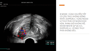
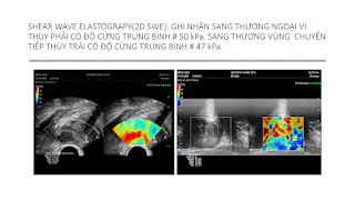
At Medic, an SWE elastography TRUS with biplane probe was done


A biopsy of prostate by via  transperineal perfomed and histopathological result is TB of prostate.


CASE 618: SMALL UTERUS LEADS TO SUSPICION TO CEREBRAL TUMOR, Dr LE DINH VINH PHUC, Dr PHAN THANH HAI, MEDIC MEDICAL CENTER, HCMC, VIETNAM

$
0
0


Female tennager 13 yo from midland region of middle Vietnam go to Medic for a checkup. 

In general ultrasound  a small uterus was detected so inducing a endocrinological problem but therer is no clue about. 


For year got pain of her right leg and foot at gym without abnormal on X-ray films. From 6 months till now 3 right hand fingers, foot and fingers are involuntary in flexion and weakness feelings. Then for later 3 months her chief complaints are plenty drinking, polyuria, headache, space out, more sleeping and drowsiness. But she got no fever nor blurred vision.


MRI brain was performed and a germinoma tumor was detected above hypophysis,hypophysis and basal ganglion on right side.


Viewing all 626 articles
Browse latest View live