Quantcast
Channel: VIETNAMESE MEDIC ULTRASOUND
Viewing all 624 articles
Browse latest View live

CASE 384: NEONATE PERINEUM TUMOR, Dr PHAN THANH HẢI, MEDIC MEDICAL CENTER, HCMC, VIETNAM

$
0
0


Neonate female 02 day-old   detected  one mass  in perineum, size of 10cm, soft in palpation [see 2 fotos].


Ultrasound  scanning of  this mass=  US1: structure of this mass is cystic  septation  with solid part.

US 2 : vessels in septation.

US 3 :sacrum and  the mass.


Sonologist  suggestion is  cystic lymphangioma.
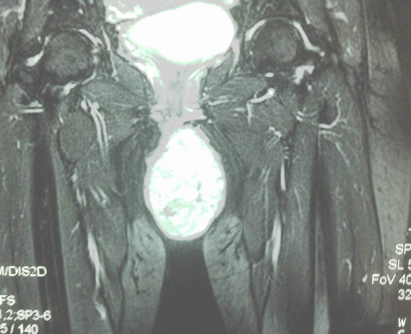
MRI  report  is   fatty  content, cystic part  not connected to spinal  canal. 


Radiologist  suggestion is  sacro-coccygeal teratoma.

Operation  removed  this mass with  solid structure and cystic part [see foto].


Report by surgeon is  mature  sacro coccygeal teratoma type 1.


MICROSCOPIC REPORT   IS MATURE  TERATOMA.


CASE 384:NEONATE PERINEUM TUMOR, Dr PHAN THANH HẢI, MEDIC MEDICAL CENTER, HCMC, VIETNAM

$
0
0


Neonate female 02 day-old   detected  one mass  in perineum, size of 10cm, soft in palpation [see 2 fotos].


Ultrasound  scanning of  this mass=  US1: structure of this mass is cystic  septation  with solid part.

US 2 : vessels in septation.

US 3 :sacrum and  the mass.


Sonologist  suggestion is  cystic lymphangioma.
MRI  report  is   fatty  content, cystic part  not connected to spinal  canal. 


Radiologist  suggestion is  sacro-coccygeal teratoma.

Operation  removed  this mass with  solid structure and cystic part [see foto].


Report by surgeon is  mature  sacro coccygeal teratoma type 1.


MICROSCOPIC REPORT   IS MATURE  TERATOMA.


CASE 385: PLANTAR, PALMAR KERATOSIS= HOWEL- EVANS SYNDROME, Dr PHAN THANH HẢI, MEDIC MEDICAL CENTER, HCMC, VIETNAM

$
0
0


Man 61 yoconsulted by  right and left plantar pain. He says it happened more one year agowith skin in foot and hand  thickening (photo)


Now he has got progressive dysphagia for one month.
Ultrasound at his neckdetected  dilatationof cervical esophagus(US 1)and  one mass  intra  esophagus at  longitudinal scanning (US 2). 


Ultrasound suggestion is  tumor of cervical esophagus.
MSCT with  oral contrast [CT 1, CT 2, CT 3] : cervical esophagus is  thickening of the wallby tumor  covered the lumen.


Endoscopy detected  exotic tumor intra esophagus lumen (endoscopic foto)  and  biosyis done.


Microscopic report is SCC (squamous cell carcinoma).


Do you see  another  case  having relation  between  palma-plantar  keratosis and esophagus cancer ?

UPDATE= HOWEL-EVANS SYNDROME.


CASE 386: ADRENAL TUMOR, Dr PHAN THANH HẢI, MEDIC MEDICAL CENTER, HCMC, VIETNAM

$
0
0


Man 44yo  high blood pressure. Ultrasound  screening of kidney detected one mass with size of  12 cm at upper pole of right kidney which was well bordered  and hyperechoic ( see  US 1)
US  2 and  US 3=  CDI  findings of  normal vascular kidney.


CT scan  with CE=  this mass is well bordered,  below liver  and pushed righ kidney down.


MRI  detected this mass more fatty structure  and  suggested that was a myolipoma of right adrenal gland.


Blood tests detected nothing abnormal.

Do you make a diagnosis  of  myolipoma for  adrenal gland?

OPERATION REMOVED  A ROUND TUMOR ( SEE MACRO).


Microscopic report   of this tumor is Myolipoma  of adrenal gland.

REFERENCE

http://downloads.hindawi.com/journals/criu/2013/789481.pdf


CASE 387: LIVER ABSCESS DUE TO FISHBONE, Dr PHAN THANH HẢI, MEDIC MEDICAL CENTER, HCMC, VIETNAM

$
0
0


Man 52 yo with pain  in liverand history of treated  liver abscess by ultrasound  guided puncture and drainage for 1 month..
Ultrasound  scan at liner=


 US 1:detected  liver  abscess  in recovery phasewith one  echo rich  body  in the boder of livercame from duodenum.


US 2: CDI no change of vascular structure of liver.


US 3 : elastoscan  showed this body had  very redcode whicn means  very hard.
CTliver  scanning  also detected  foreignbody ( fb) intra liverand one site was intra duodenum wall.


Emergency  endoscopy  cannot detect  this fb.
Laparoscopy  removed the fish bone  with 3.5 cm length (  foto)


Conclusion: liver abscess  due to  fishbone  migrated from duodenum to liver.
Reference:

Fish Bone Penetration of the Duodenum: A Rare Cause of Liver Abscess


CASE 363: MURPHY'S SIGN POSITIVE, Dr PHAN THANH HẢI, MEDIC MEDICAL CENTER, HCMC, VIETNAM

$
0
0


Woman  32 yo,  3 days ago, fever and pain at  right  upper quadrand of abdomen with  MURPHY SIGN  POSITIVE  in clinical palpation.
Report of ultrasound in emergency from  a province hospital   was cholecystitis necrosis and peritonitis ( US picture).

At MEDIC, reviewed ultrasound shows US 1: CDI revealed big gallbladder and edema of the wall, no stone, no perforation. CBD is  no dilatation, no hypervascular.


US 2: fluid collecting in Morrison’s space extending to right iliac fossa.


US 3:normal scanning  at pancreas area.


Patient reports painful in pressing of ultrasound probe over gallbladder area .
Sonologist  suggested  edema of the gallbladder wall  and ascites maybe  due to hemorragic fever reaction.
Blood tests  confirmed  low WBC, low platelets, and Dengue test  IgG positive.


Based on  ultrasound  picture and  blood tests, diagnosis was infected Dengue; gallbladder edema only due to reaction. And the management for the case  is  medical follow-up in progress of disease.
Reference:
Acute Acalculous Cholescystitis and Ascites [Dengue Fever stage III]
Hình ảnh siêu âm sốt xuất huyết Dengue

CASE 364: LUNG LOOKED LIKE LIVER, Dr PHAN THANH HẢI, MEDIC MEDICAL CENTER, HCMC, VIETNAM

$
0
0


Woman 62 yo, cough and dyspnea, weakness of left side of  her body  2 weeks ago.
Chest XRay  first.( see pleural effusion  at right lung).


Ultrasound of  thorax:
US1=liver normal with mass  at  lower portion of right lung


US 2=liver and right lung  looked like liver structure (hepatization).


US 3= scan at right thorax: pleural effusion and lung solid mass.


US 4=  with 10MHz linear probe  looking of visceral layer of pleural membrane having  irregular nodular mass.


US 5 =  this lung mass is hard  like liver.


US 6= very low vascular supplying.


CT scan of lung  non CE.: CT1=cross section,  CT2 = frontal view,  CT 3= many nodular  metastasis at right and left lung.


CT4=  brain scan with suggestion of metastasis at right brain..
Punction of pleural space removing yellow fluid ( foto).


Analysis of fluid = ADA  very low, ruling out lung tuberculosis.

Do you  thing this case  is lung cancer metastasis to the brain? 

REFERENCE:
Ultrasound detection of Lung Hepatization

CASE 365: MULTIPLE INTRAMUSCULAR TUMORS, Dr PHAN THANH HẢI, MEDIC MEDICAL CENTER, HCMC, VIETNAM.

$
0
0


Woman  60 yo being treated   lymphoma large B cell  stage IV by chemotherapy for 5 months.

One week ago she herself detected  many  subcutaneous nodules palpable  at  forearm right and left, neck and  right parotid area, no painful.
 ULTRASOUND=
US 1=tumor  intramuscular right  forearm, round  border, very  low echo density.


US 2=cross-section, lesion at forearm.

US 3=CDI  Doppler vascular  structureof this mass, hypervascular.


US 4=longitudinal scanning  with  CDI.


US 5=CDI with PW,   RI= 0,70.


US 6 = small intramuscular nodule  at posterior of  neck.


US 7= SWE of mass in right  parotid.


Do you thing  it is lymphoma  in muscle?  
Biopsy of this mass  is large  B cell lymphoma, same as  result pre-treatment.


Conclusion: LYMPHOMA  LARGE B CELL  AT THE DIFFUSE STAGE  CAN MAKE  MULTIPLE NODULES  IN MUSCLES.
Reference:


CASE 388: TESTIS TUMOR, Dr PHAN THANH HẢI, MEDIC MEDICAL CENTER, HCMC, VIETNAM

$
0
0


Boy 11 yowith left  scrotum is bigger than right one, he went through an  operation for  left inguinal hernia  2 years before, now  no pain, no fever.
US of scrotum detected hydrovaginalis and a small focal intratestis, size  of 0,8 cm, cystic calcification  in septation ( US 1, 2, 3), hypovascular 


and very hard  in elastography( US 4).


MRI reported a cystic tumor with calcificationwith  size of 1 cm.


All  AFP, HCG, testosterone  or  corticoid of blood tests arenegative.

Operation  for biopsy: macroscopic tumor is intra testis,  not  invasion to tunica vaginalis; tumor is white structure  like caseum.


 Wait for  microscopic report.

CASE 389: ECTOPIC APPENDICITIS, Dr PHAN THANH HẢI, MEDIC MEDICAL CENTER, HCMC, VIETNAM

$
0
0

Man 35yo, onset  epigastric pain  treated  as gastritis for one week, fever.
Ultrasound of   abdomen= pain  at  upper  left  abdomen quadrant (US 1  probe put over  pain point).


US  2= mass  rounding as  an abscess, CDI: no  more vascular  supply.
US 3 (with linear probe)  intra abscess the linear structure  look like  appendix).
US 4 =  cross section.


Emergency CT with CE=   mass  wall-off  by  small intestine and  great omentum  as an abscess  ( CT1, CT2).


Blood test  WBS = high 14k , with neutro 9.9,  CRP  38.3ng/dl.
Preoperative diagnostic is  intra abdomen abscess  due to ectopic appendicitis.
Laparoscopic view = the pus goes out  from this mass  and open operation removed appendiceal partial necrosis and  mobile coecum.


Conclusion=  appendiceal abscess in ectopic position at left abdomen site.

CASE 390: P-HCC, Dr PHAN THANH HẢI, MEDIC MEDICAL CENTER, HCMC, VIETNAM

$
0
0
MAN 56 YO REPORTED  HIS RUQ  SWOLLEN  SLOWLY FOR  6 MONTHS , NO DISTURB GI TRACT
IN DIGESTION.
ULTRASOUND ABDOMEN:
US 1:LIVER  AND TUMOR  NEARBY ECHOGENEICITY IS DIFFERENT.


US 2:LONGITUDINAL SCAN,TUMOR AND LIVER BORDER ARE NOT CLEAR.


US 3: STRUCTURE OF THIS TUMOR IS SOLID, HYPOVASCULAR.


US 4: CROSS SECTION, TUMOR IS INTRA ABDOMEN, AT RIGHT  SITE OF AORTA.


MSCT WITH CE:
CT 1( 4 PICTURES): SAGITTAL VIEW, FRONTAL VIEW ,CROSS.. VASCULAR SUPPLY 
OF THIS TUMOR IS FROM LIVER.


CT1:TUMOR IS  RELATED WITH  R/LIVER,   PEDUNCULATED, VASCULAR SUPPLY FROM LIVER.


CT2:TUMOR IS MULTINODULAR, CE  IS  IN BORDER OF TUMOR.


LAB BLOOD TESTS  =  HBV POSITIVE, AFP=651.8 ng/mL.

SUMMARY=   PRE OP IS SUSPECTED HCC, BUT IT  HAD PEDUNCLE  COME FROM RIGHT LIVER. LAPAROTOMY REMOVED BIG TUMOR  FROM THE RIGHT  LIVER.

CASE 391: DOUBLE BREAST TUMOR, Dr PHAN THANH HẢI, MEDIC MEDICAL CENTER, HCMC, VIETNAM

$
0
0


Woman 53 yo, for one month  detected  her breast distention both 2 sites and  pain. In clinical examination 2 breast are hot and hard.
Ultrasound  (US 1), right breast had some echo poor focal lesions, 2-3 cm, non compressible.


US 2: same  structure at the left  breast.


US 3=


US 4: color Doppler  hypovascular.


Mammo Xray  =  very dense breast (MM1, 2).


MRI with gado=  breast are filling by  hyperintense mass  with gado enhancement.


Bood tests= WBC  normal, beta microglobuline raised 2,200 UI (n=2,100 UI)
Biopsy was done  by core biopsy  and  IHC staining  report is  lymphoma large B cell.


Conclusion= Lymphoma is most common appearance of  2 breast simultaneous infiltration.

REFERENCE

DOWNLOAD PRIMARY BREAST LYMPHOMA


CASE 392: PERFORATED SEAL-OFF DUODENUM, Dr LÊ TỰ PHÚC-Dr PHAN THANH HẢI. MEDIC MEDICAL CENTER, HCMC, VIETNAM.

$
0
0


"A 37-year-old male presented in our hospital with moderate epigastric pain for three months. Around one week before examination, the pain became more severe, but he didn’t recognize a suddenly pain. Clinical examination revealed no muscle guarding and rebound tenderness.

Abdominal ultrasound images showed unconcentric wall thickening of the gallbladder. Beside the more thickening wall of gallbladder, a hyperechoic of air collection was found. This air collection was continuous with small hyperechoic air spots inside duodenum. A perforated duodenal ulcer with air leakage was suspected.


CT-Scan confirmed air collection beside a thickening gallbladder wall.


Blood test indicated and raised of white blood cells (10,350 / mL) with low level of CRP (0.9 mg/L) and possitive Helicobacter Pylori test.

Without surgery, the patient pain released and the air collection was disappear in ultrasound and CT-Scan images for one month follow-up. Gastroduodenal endoscopy showed a healing ulcer in the anterial wall of duodenum. White blood cell count returned to normal."


This is a case of perforated seal-off duodenum revealed by ultrasound and confirmed by CT-scan later and successfully management without surgery.

CASE 393: SPLEEN TUMOR, Dr PHAN THANH HẢI, MEDIC MEDICAL CENTER, HCMC, VIETNAM

$
0
0

Woman 26 yo with no clinical symptom. Ultrasound screening detected a spleen mass
US 1= longitudinal scan of this mass size of 6.0cm at lower pole of spleen, hypoechoic, well bordered.

US 2=cross-sectional view of mass.


US 3=CDI of this mass with vascular bending sign and, ( US 4) structure inside hypervascular.


Blood tests are normal.
CT with CE:CT 1 non CE , CT2 CE, delay phase with central mass lower perfusion.


MRI with gado: this tumor is well bordered, peripheral enhanced and central hypoperfusion at the late phase.


LAPAROSCOPY  FOR  SPLENECTOMY  . SURGEON REPORTED  THIS TUMOR IS INTRA SPLEEN AND ITS COLOR  LOOKS LIKE SPLEEN TISSUE ( SEE  SPECIMEN).HE SAID IT MAY BE  HEMANGIOMA.


CASE 394: COLON TUBERCULOSIS, Dr PHAN THANH HẢI, MEDIC MEDICAL CENTER, HCMC, VIETNAM

$
0
0

Man 43 yo with epigastric pain  crisis and  gastroendoscopy showed  gastritis.
Ultrasound  detected  one mass  like target  with  thickening of the  wall of colon (see  US 1=csoss-section  colon over  right kidney);   US  2  with linear  probe= colon wall is thickening; US 3, US 4 =  longitudinal scan).


MSCT of abdomen with CE revealed  thickening of  ascending colon wall  (CT1,  CT2).


Chest X-rays  before endoscopy  detected  infiltration of  left upper lung.


Colonoscopy reported the mass in right colon, nodular ( see foto) biopsy. 


Report of endoscopist is colon cancer.
Microscopic report  is colon tuberculosis.
Conclusion = this  case  represented colicky pain at epigastric region but  ultrasound and CT  suggested  colon cancer, same as colonoscopy, but  microscopic is tuberculosis.of colon and left lung.

REFERENCE:


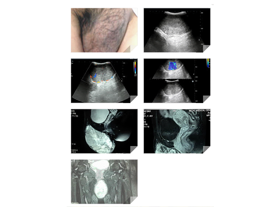
CASE 395: MAJOR LABIA TUMOR, Dr PHAN THANH HẢI, MEDIC MEDICAL CENTER, HCMC, VIETNAM

$
0
0


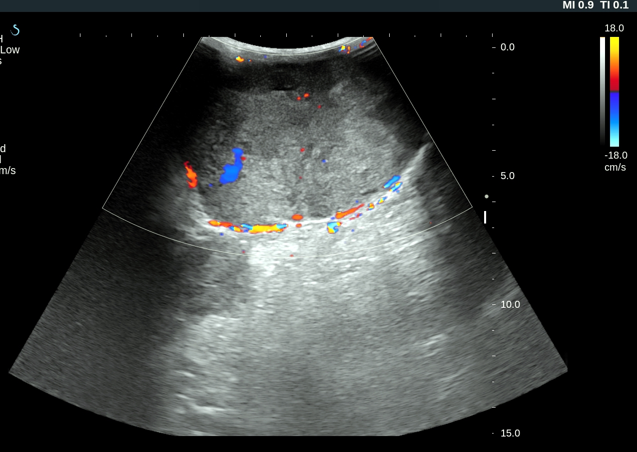
WOMAN 45 YO, SAYS  THIS VULVA MASS  IN  SLOW GROWING 6 MONTHS AGO, WITHOUT  PAIN ( FOTO).


ULTRASOUND OF THIS TUMOR REVEALED SOLID, FIRM,   SIZE OF 10CM.
 US 1:WITH  B MODE,    US 2 WITH  CDI : HYPOVASCULAR SOLID TUMOR /  US 3=ELASTOULTRASOUND OF THIS TUMOR IS HARD STRUCTURE.


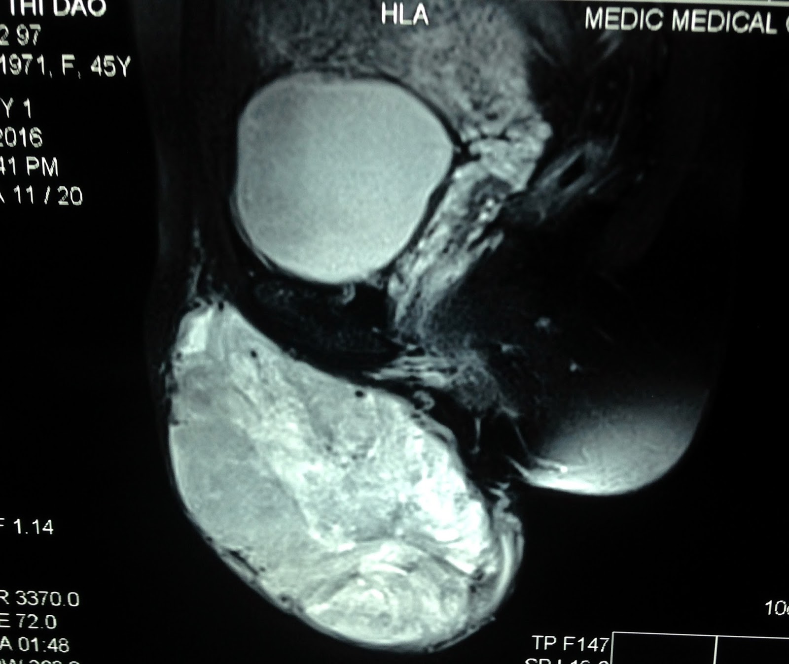
MRI 1 SCAN = SAGITTAL SECTION OF THIS TUMOR
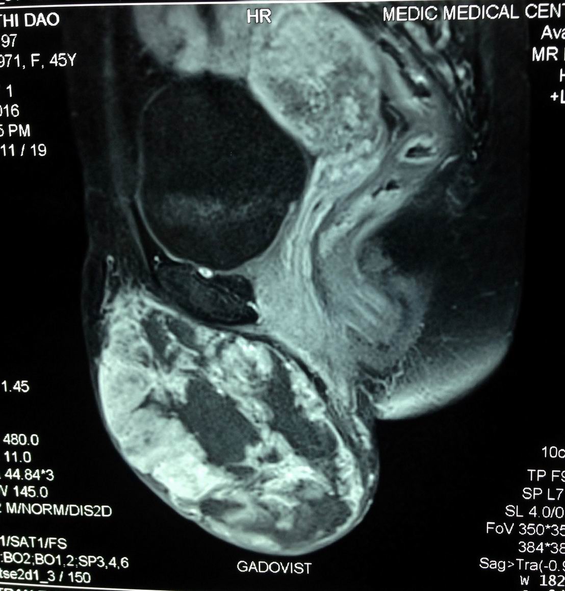
MRI 2 WITH  GADO  CE= IN LATE PHASE, LOOK LIKE  A CYSTIC DEGENERATION.
 MRI 3 = FRONTAL  SECTION OF THIS MASS  FROM LEFT MAJOR LABIA,  PEDUNCULATED  LOOK  LIKE SCROTUM IN MALE PATIENT.


Core  biopsy reported  microscopic with  immmunochemistry staining is  glomus tumor.


CASE 396: PARATHYROID CYST, Dr PHAN THANH HẢI, Dr NẠI HƯƠNG THOANG, Dr VŨ TU THÂN

$
0
0


Woman 43 yo with  sorethroat  and cough, sputum bleeding.
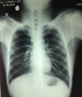
Chest X-Rays for  screening:   no  chest lesion ( see foto chest X-Rays).


Ultrasound of  the neck: normal thyroid  but detected a cyst at lower pole of thyroid gland, size of 5-6 cm, monocystic  prolonged to retrosternum.
US 1,US 2 ( CDI),   US 3    pretrachea longitudinal scanning.


MSCT CE of  the neck: CT 1=frontal viewing,  well bordered cyst, CT 2: sagittal view..
C T 3. Cross-section= retrosternum tumor.


Ultrasound guide punction  of this cyst removing 10ml clear fluid.
What do you need to study in this fluid?

fluid analysis report: high PTH  78.69pg/ml  and in the blood PTH is 47pg/mL.
The final diagnosis is  non functional PARATHYROID  CYST.
  
Reference


CASE 397: NERVE TUMOR, Dr PHAN THANH HẢI, Dr NGUYỄN NGHIỆP VĂN, MEDIC MEDICAL CENTER, HCMC, VIETNAM.

$
0
0


Boy 17yo with pain at anterior brachial both 2 sides. Clinical palpation revealed many small subcutaneous nodules, size as a pepper nut, painful at palpation ; and swelling neck (see chest XRays).


Ultrasound scanning of the antebrachial areas detected many small ellypsoid nodules along the arterial way looked like the nerve ( US 1, US 2: longitudinal scan and cross-section).


US 3, US 4 scan at the carotid detected hypoechoic mass along the carotid sites  which belong to vagus nerves 2 sites.


US 5, US 6 scan at left carotid= big size vagus nerve, hypoechoic #1.5 cm in diameter at cross section.


MRI of the neck and thorax= MRI 1, MRI 2: hypertrophic nodular vagus nervi go to  mediastinum.


MRI 3: lateral view of the neck; MRI 4 = cross section of vagus nerve at the neck in relation with carotid artery.


MRI 5: hypertrophic intercostal nerve at thorax.

Biopsy of  the subcutaneous nodule at forearm  reported  neurofibroma in microscopic result.


CASE 398: ECTOPIC THYROID GLAND, Dr PHAN THANH HẢI, Dr TRẦN THỊ BẢO CHÂU, Dr NGUYỄN THIỆN CHÂU

$
0
0


Woman 48 yo, PARA 2002, in general check-up, sonologist  detected no thyroid gland at normal location (US 1).


Blood tests reported  TSH= 11.47 microIU/mL (n= 0.3-5 microIU/mL),  Free T4= 0.985 ng/dL ( n= 0.7-1.85 ng/dL).
Ultrasound  again..detected  one mass  at  basal tongue,  size of 3x4 cm, hypoechoic,  hypovascular  (US 3, US 4).


MSCT with CE  detected  intralingual  mass which is  very quick  enhanced of  CE, size of  4 cm (CT1, CT 2, CT3).


CTA  detected  no thyroid arteries both  superior or inferior branches from the CCA and  ECA ( CT 4)


Conclusion=  Ectopic thyroid gland  intra basal lingual type.

CASE 399: HCC GOING TO HEART, Dr PHAN THANH HẢI- Dr NGUYỄN TUẤN VŨ, MEDIC MEDICAL CENTER, HCMC, VIETNAM

$
0
0


MAN 66 yo with CHEST PAIN and DYSPNEA. EMERGENCY ECHOCARDIOGRAPHY DETECTED  INTRA CARDIAC MASS, LOOKED LIKE THROMBUS.
ULTRASOUND SCAN of LIVER DETECTED a LIVER MASS SIZE of 4cm and DILATED 2cm in DIAMETER and OBSTRUCTED by A HYPOECHOIC MASS ( US 1, US 2).


MSCT with CE DETECTED LIVER MASS and TUMOR INVASION TO HEPATIC VEIN TO IVC and GOING to RIGHT ATRIUM and FILLING DEFECTED at PULMONARY ARTERY (CT1, CT2).


CT3 (section) SHOWED LIVER MASS, RIGHT ATRIUM MASS and INTRA LEFT VENTRICULAR MASS.


BLOOD TEST = HCV POSITIVE; WAKO TEST = TRIPLE POSITIVE.

Viewing all 624 articles
Browse latest View live