Quantcast
Channel: VIETNAMESE MEDIC ULTRASOUND
Viewing all 624 articles
Browse latest View live

CASE 326: INTUSSUSCEPTION of COLON INDUCED by POLYP, Dr LÊ TỰ PHÚC, MEDIC MEDICAL CENTER, HCMC, VIETNAM

$
0
0
FOR PICTURES PLS CONNECT TO 3G /DOWNLOAD THE LINK

A 4 years-old boy presented to Medic Center with one year history of weakness, fatigue, lethargy, pale skin and less active. No recognition of other symptoms such as vomiting, abdominal pain or bloody stools. Patient was done blood test and abdominal ultrasound. 
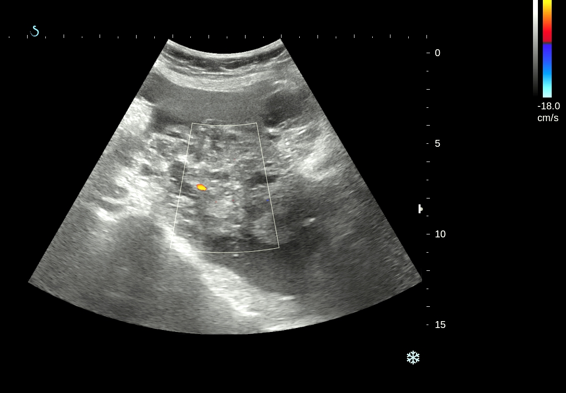
Abdominal ultrasound detected colo-colonic intussusception in the right upper quadrant with concentric rings sign in transverse scan and "hay fork" sign in long axis scan. Located adjacent the intussusception show an isoechoic to hypoechoic solid mass, well defined oval, 30 mm in diameter, hypervascular in the hilus of the mass. Those blood vessels were continuing with the blood vessels from central portion of the intussusception. Sonologist suspected a intussusception of the ascending colon secondary to a  polyp. 


Laboratory investigations showed the reduction of Hemoglobin: 6.5 g / dl.


The patient was transferred to the hospital Nhi Dong 2. He had positive fecal occult blood test. Colonoscopy showed a polyp of ascending colon. 


A surgery was then obtained 2 weeks later.


 Surgical results confirmed polyp of the ascending colon which pathology result is tubular polyp.

CASE 327: INTRATHORACIC THYROID TUMOR, Dr PHAN THANH HẢI, MEDIC MEDICAL CENTER, HCMC, VIETNAM

$
0
0
FOR PICTURES PLS CONNECT TO 3G/ DOWNLOAD THE LINK

Woman 37 yo with history of  sore throat and  changed  speaking.
Chest X-Ray detected  one  mass at upper pole of right lung (chest x-ray film).


Transthoracic ultrasound  of this mass revealed  a solid  hypovascular mass, size of 10 cm, no moving  with  respiration. Thyroid  ultrasound  scan  was normal  but the right lobe was  smaller  than left one.


MSCT  angio in  cervico-thoracic region showed mass in connecting with right  thyroid lobe.
Main vascular supplying for this mass was from branch of  inferior thyroid artery  (see CTA).


Operation for  removing of  one round mass connected with  right lobe of thyroid, its
structure was inhomogeneous  and pathology report was  colloidal  thyroid goiter.


Conclusion: this case  was  an intrathoracic goiter while on chest X-Ray film  looked like  lung tumor.
Reference:
Case from Mayo Clinics 


CASE 328: DISSECTING SUPERIOR MESENTERIC ARTERY, Dr LÊ VĂN TÀI, MEDIC MEDICAL CENTER, HCMC, VIETNAM

$
0
0
FOR PICTURES PLS CONNECT TO 3G/DOWNLOAD THE LINK


Male patient  50yo, old patient of Medic Center with history HTA and prolonged epigastric pain , HP infected.

Epigastric pain was getting worse for one month, he was admitted by province hospital for 10 days and had been treated as gastritis, but continued gastric pain out of hospital for 20 days. So coming back to Medic Center.


Ultrasound at Medic Center revealed dissecting SMA at its origin, d=12mm, raising diameter and flapping moving inside in TM mode, aliasing in color mode,  and confirmed it later by MSCT.


REFERENCES:


CASE 329: MEDIASTINAL TUMOR, Dr PHAN THANH HẢI, MEDIC MEDICAL CENTER, HCMC, VIETNAM

$
0
0
FOR PICTURES PLS CONNECT TO 3G /DOWNLOAD THE LINK


Man 28yo, one week ago getting of chest pain, fever and cough. Chest X-Rays (image 1)  showed a  mass  at  left  lung which was thought to be a left anterior mediastinal tumor.


Transthoracic  ultrasound reported  a solid mass, size of  6.7 cm, hypovascular, covered  left anterior mediastinum with  pericardial effusion (ultrasound images 1, 2, 3, 4).


MSCT with CE made diagnosis with a left anterior mediastinal tumor like thymoma.


Blood test made attention to  AFP very high value (lab report).


Clinical status  and  XRays film , US,  CT and  lab reports suggested a  teratocarcinoma (seminoma).
Core biopsy of  this tumor report was an  undifferentiated adenocarcinoma.


The  patient is  waiting for  operation.

CASE 330 : SMALL HCC, Dr PHAN THANH HẢI, MEDIC MEDICAL CENTER, HCMC, VIETNAM

$
0
0
FOR PICTURES PLS CONNECT TO 3G/DOWNLOAD THE LINK

case-330-small-hcc-

Man  66 yo with history  no infected  HBV, HCV but  high values of liver enzymes for a long time.

Ultrasound of liver showed fatty infiltrating in liver.

Blood test of HCC RISK  positive  with  .AFP : 33.6 ng/ml   AFP-L3%: 62,4% and DCP: 21mAU/mL.


MRI of liver  with PRIMOVIST and DWI detected a  small focal lesion, size  of 0.8cm at the liver border in  segment 6,  very  bright  in DWI  and captured  and washed out  Primovist like a HCC.


Diagnosis  was made for a small HCC,  wait for operation.

CASE 331: MASS INSIDE STOMACH, Dr PHAN THANH HẢI, MEDIC MEDICAL CENTER, HCMC, VIETNAM.

$
0
0


FOR PICTURES PLS CONNECT 3G / DOWNLOAD THE LINK


Child 5 yo long history of epigastric pain and anorexia treated  as  HP  infected gastritis.

Ultrasound detected intragastric mass  with  large UMBRELLA SIGN  [showdown  like the  sunshine over the umbrella] which  covered  more  50%  of  lumen of stomach ( see US pictures 1, 2, 3).


MSCT of  abdomen revealed this mass  inside  stomach,  multilayer  structure, not  developmental from the wall of  stomach.


Based on ultrasound  and abdomen CT, suggestion  of radiologist  is  gastric trichobezoar.

What is your  suggestion, endoscopy  or  operation for removing the mass inside stomach?
Operation of gastrostomy removed the hair mass.


Reference:
Umbrella's sign of ultrasound.
https://www.google.com/?gfe_rd=cr&ei=g5_tU8msNeuJ8Qfjl4HADg&gws_rd=ssl#q=umbrella%27sign+ultrasound&imgrc=iff-XjzsO-ZxoM%3A

CASE 332 : PERITONEAL CARCINOMATOSIS, Dr PHAN THANH HẢI, MEDIC MEDICAL CENTER, HCMC, VIETNAM.

$
0
0
FOR PICTURES PLS CONNECT TO 3G /DOWNLOAD THE LINK


Woman 68 yo with abdomen distension and pain.
Ultrasound  scan detected  ascites  with   pelvic mass ( US images) suspected  peritoneal carcinomatosis.


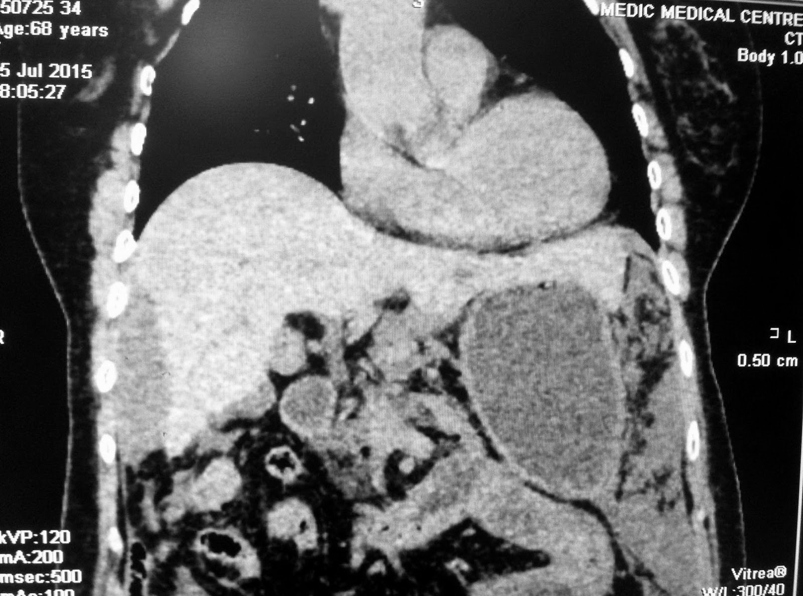
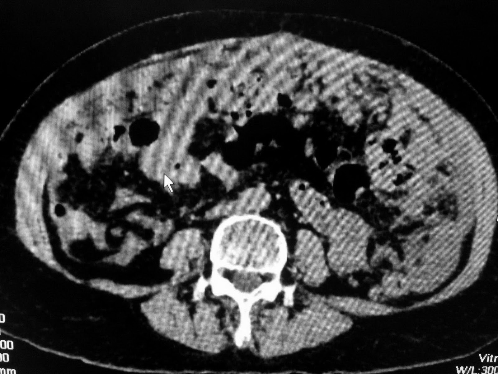
CT  scan of abdomen with CE with  radiologist  report  was   peritoneal carcinomatosis,
unknown  primary cancer (CUP).


Blood tests:   raised CEA and  CA-125 markers.


Abdomen tap removed gelatineous fluid that analysis of  this fluid no cancer cell revealed.


Laparoscopy biopsy of  peritoneal  vegetation reported  mucinous carcinoma.

Discussion:  In this case  of  68 yo female ascites  like  jelly, CT and ultrasound  cannot detected ovary tumor. Blood tests  pointed  CEA  very high in comparison to CA-125.  The most common cause is  from rupture of appendiceal.carcinoma and  spreading  intraperitoneum.


Ref..


CASE 333: LUNG MASS, Dr PHAN THANH HẢI, MEDIC MEDICAL CENTER, HCMC, VIETNAM

$
0
0
FOR PICTURES PLS CONNECT TO 3G / DOWNLOAD THE LINK


Man 52 yo, fever  unknown  origine  for 3 months, blood tests: nothing abnormal detected.
MSCT scan of full bodydetected  a small nodule on right lung,  size of 1 cmwithsome  pericarena lymphnodes enhanced with  CEand  one subcutaneous mass of 3 cm in the  back of  left  neck (CT lung images).


Ultrasound of this mass  revealed round border, very hypoechoic, nonvascular filling intramass, no posterior enhancement, no sister mass together ( see 3 US images and  video clip).


Biopsy was done for this mass and microscopy result was  adenocarcinoma  metastasis  from the lung.


Discussion: Clinical onset is fever unknown origine, CT lung detected small spicular nodule , pericarena nodes and the patient himself detected one subcutaneous mass at posterior of his left neck; biopsy of this mass made diagnosis of metastasis from lung cancer  which is small cell lung cancer.

Conclusion= Small  lung cancer  metastasis to skin  and  paraneoplasic  fever.
Reference: Case in NEJM.


CASE 334: MULTIPLE HEPATIC TUMORS: Dr PHAN THANH HẢI, MEDIC MEDICAL CENTER, HCMC, VIETNAM.

$
0
0
FOR PICTURES PLS CONNECT TO 3G / DOWNLOAD THE LINK
case-334-multiple-hepatic-tumors-

WOMAN 43YO, CHRONIC INFECTED HBV, PAIN AT  LIVER.
ULTRASOUND   DETECTED  INTRA HEPATIC   MULTIPLE FOCAL LESIONS WITH SUGGESTION OF  LIVER HEMANGIOMA (US1, US 2).


REPORT OF MSCT WITH CE   WAS    MADE  DIAGNOSIS OF   MULTIPLE  FOCAL HCC (  CT 1, CT 2).


MRI WITH  PRIMOVIST ALSO   WAS   DIFFUSE   HCC.


BLOOD TEST OF   WAKO  WERE  NEGATIVE,  CEA AND CA 19-9   WERE  IN  NORMAL  LEVEL.
FIBROTEST  WAS  SCORED  F2.


WHAT IS  YOUR   SUGGESTION FOR DIAGNOSIS?.


CASE 335: HEMOPERITONEUM, Dr PHAN THANH HẢI, MEDIC MEDICAL CENTER, HCMC, VIETNAM.

$
0
0
FOR PICTURES PLS CONNECT TO 3 G / DOWNLOAD THE LINK


WOMAN 33 YO, ACUTE   LEFT PELVIC PAIN, ULTRASOUND  EMERGENCY  SHOWED NORMAL SIZE OF UTERUS, ENDOMETRIUM  IN MIDDLE OF MENSTRUAL CYCLE, AND HAVING A LOT OF FLUID AND  BLOOD ARROUND  UTERUS TO MORRISSON'S  SPACE  OF ABDOMEN.
IT WAS A CASE OF HEMOPERITONEUM.

ULTRASOUND  DETECTED  ONE ROUND  MASS  AT LEFT  PELVIS WHICH WAS  SUSPECTED AN  OVARY CYST WITH SIZE OF 4 CM.


PUNCTION OF ABDOMEN  AT  RIGHT ILIAC FOSSA  REMOVED OLD  BLOOD.
BLOOD TESTS  WERE NORMAL  BETA HCG,  Hct   21%, NORMAL AMYLASEMIA.

MSCT  WITH CE  DETECTED  2 MASSES  BOTH 2 SIDES RIGHT AND LEFT  UTERUS; THE LEFT ONE  WAS  VERY QUICK CE ENHANCED  IN COMPARISON TO THE RIGHT  ONE  NON-CE ENHANCE.


Patient was  in admission of emergency of  surgery hospital.
After receiving of  2 units of blood transfusion, the vital status was stable.
Follow- up by  ultrasound one week later  the  blood clot  in pelvis was not rising  but not dissolved. Laparoscopic surgery  for  washer  this blood  and  detected  right ovary was normal, while  left ovary  had  ruptured one cyst but  already  stop bleeding.

Report of surgeon was rupture  of luteinic corpus  with blood loss more than 2,000 ml-- a  severe case of internal bleeding.

CASE 336:2 year Abscess in Leg Muscle due to Missed Foreign Body, Dr LÊ THANH LIÊM, Dr LÊ THÔNG LƯU, Dr PHAN THANH HẢI, MEDIC MEDICAL CENTER, HCMC, VIETNAM

CASE 337: BILATERAL BREAST TUMOR, Dr PHAN THANH HẢI, MEDIC, MEDICAL CENTER, HCMC, VIETNAM

$
0
0
FOR PICTURES PLS CONNECT TO 3 G/ DOWNLOAD THE LINK


WOMAN  30 YO, FOR CHECK-UP MAMMOGRAPHY  DETECTED  CALCIFICATION MASS  IN RIGHT AND LEFT  BREAST AND RADIOLOGIST SUGGESTED  BIRADS 4 BREAST TUMORS ( SEE  XRAY MAMMO ).


ULTRASOUND  OF RIGHT BREAST DETECTED  ONE HARD MASS,  IRREGULAR BORDER   WITH VERY STRONG  POSTERIOR ATTENUATION  WITHOUT AXILLARY LYMPH NODE ( SEE  US 1, 2, 3).


ULTRASOUND SCANNING  OF LEFT BREAST  ALSO  HAD  SAME  TEXTURE  MASS WITH  IRREGULAR BORDER   WITH  HIGH  ATTENUATION.


SONOLOGIST    REPORT   WAS  ALSO BILATERAL BREAST  TUMOR IN SUGGESTION MALIGNANT  BIRADS 4.

ULTRASOUND GUIDED CORE  BIOPSY of RIGHT AND LEFT  BREAST TUMORS,   RESULT WERE FIBROADENOSIS CALCIFICATION.


REFERENCE:


CASE 338: THYROID CANCER, Dr PHAN THANH HẢI, MEDIC MEDICAL CENTER, HCMC, VIETNAM.

$
0
0
FOR PICTURES PLS CONNECT TO 3 G / DOWNLOAD THE LINK

case-338-thyroid-cancer


Man 30yo, in general check-up,  ultrasound detected  thyroid tumor  at right and left  lobes.
US1 scan at   right lobe, small nodule 1cm diameter,  hyperechoic due to  calcification.


US 2 scan at  left  lobe,    round border tumor, 4 cm with many white calcification spots.


US 3 & US 4:  CDI of  left  thyroid tumor, hypervascular.


US 5  elasto scan of right  tumor was very hard.


US 6 elastoscan with Q box score,  tumor in comparison to  normal thyroid tissue.


No detection of  regional lymph nodes.
Report  by  sonologist   was suspected  thyroid carcinoma, THYRADS IV, and FNAC of  the left tumor was  PAPILLARY  CARCINOMA.


DISCUSSION: B MODE  SCAN  THYROID TUMOR   WITH MANY  WHITE SPOTS  WITHOUT  SHADOWING, IT IS   MICROCALCIFICATION NAMED   PSAMMOMA BODY..WHICH  IS  TYPICAL  OF  PAPILLARY THYROID CARCINOMA.

ELASTOSCAN THIS TUMOR   WITH  QUANTITATIVE Q-BOX  IS 99.5 kPa  IN COMPARISON WITH   NORMAL THYROID  GLAND IS  12.2 kPa.
ELASTOSCAN  IS NEW  TECHNOLOGY  FOR  DETECTION  THYROID  CANCER.


REFERENCE


CASE 339: INTRAMUSCULAR TUMOR, Dr PHAN THANH HẢI, MEDIC MEDICAL CENTER, HCMC, VIETNAM.

$
0
0
FOR  PICTURES PLS CONNECT TO 3 G / DOWNLOAD THE LINK


Man 51 yo  detected  himself  swollen at  left  scapula region, no pain for 2 years (photo). 


The tumor  was  underskin and ovoid shape.
Ultrasound  of this mass was  localized   in  trapezius muscle, well bordered,  size of 10cm. CDI  no abnormal blood  flow. Elasto scan was  slow  kPa (7.7kPa) in  comparison to muscle 22.9 kPa.


MRI  scan= MRI 1,tumor  well bordered with  density as  fatty tissue, MRI 2, MRI 3).


Operation  was done  for removing of  the tumor.
Microscopic report of this tumor is LIPOMA.


CASE 340 : UMBILICAL TUMOR, Dr PHAN THANH HẢI, MEDIC MEDICAL CENTER, HCMC, VIETNAM

$
0
0
FOR PICTURES PLS CONNECT TO 3G / DOWNLOAD THE LINK

Woman 45 yo, PARA 2002, normal spontaneous vaginal deliveries,no cesarean operation  nor history of hormonal contraception. 

2 years ago  she  detected  her umbilicus swelling some days before her menses and continuous pain  during  the entire of her period in some times bleeding.
In clinical  examination  the umbilicus  deformed  by one mass which were bluish-black, hard and not  hot (see 2 photos).


Ultrasound findings of this mass=
US1: this mass was well bordered, localized  intra cavity of  navel. Structure of mass was solid, size of  2.68 cm.


US 2:  in CDI, vascular  supplying was from peripheral part of mass.


US 3:  in elastography:  this mass was hard in comparison to muscle.


US 4 : uterus and pelvis were  nothing  abnormal.


What is your  suggestion for diagnosing of this case.

CASE 341: THYROID TOXIC ADENOMA, Dr LÊ TỰ PHÚC, MEDIC MEDICAL CENTER, HCMC, VIETNAM

$
0
0

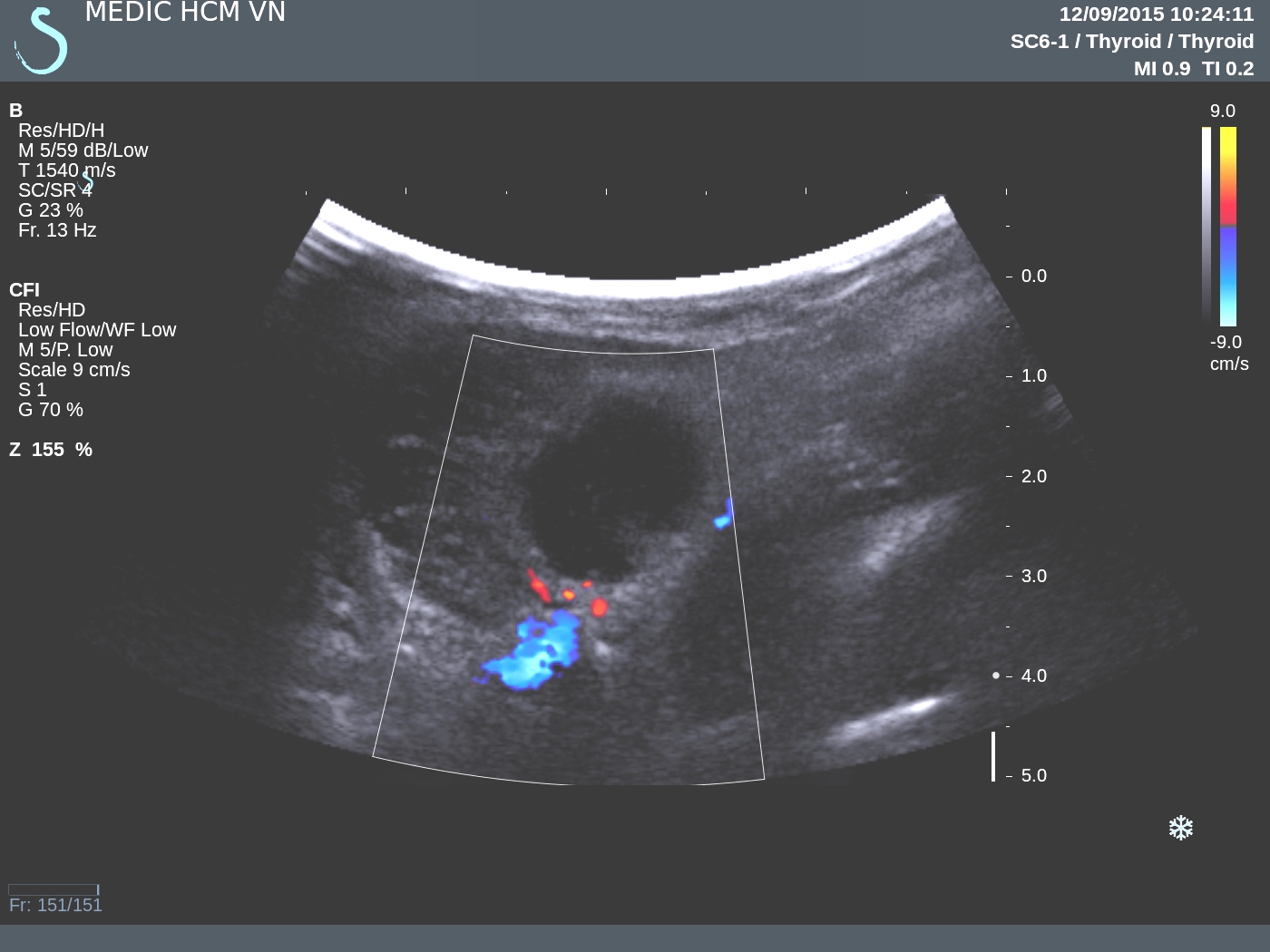
A 41 yo male patient with chronic fatigue syndrome and  nervousness, irritability; sometimes  muscle weakness and hand tremor  for three months and getting severe in ten days. Want to check up his liver and nervous system. 

Abdominal ultrasound revealed nothing abnormal. Because of his symptoms, sonologist also perfomed a thyroid ultrasound.

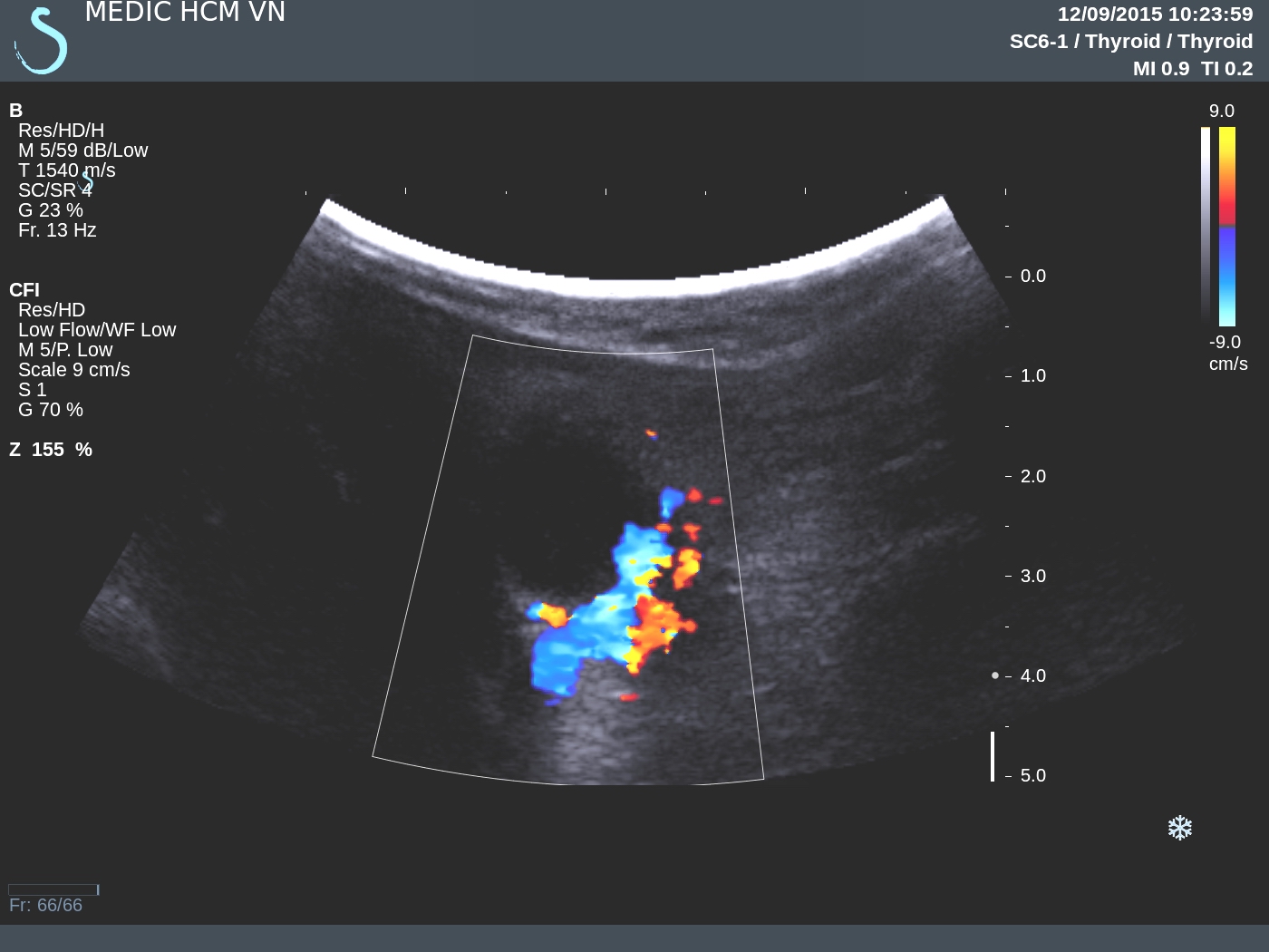
Thyroid ultrasound showed that  right lobe and  upper portion of  left lobe were normal in size with smooth margin and homogeneous echotexture, normal blood flow in Doppler ultrasound.

But  lower portion of the left lobe had a 5 cm, mixed cystic-solid nodule with hypervascular, isoechoic in peripheric part and nonvascular cystic degeneration in center part of tumor.


On Doppler US,  inferior thyroid artery showed  peak systolic velocity in  left lobe is 122.7 cm/s, five times more than one of right lobe 24.3 cm/s. So,  sonologist suspected  nodule in  lower left lobe maybe a toxic thyroid adenoma, which is cause hyperthyroidism.


Blood tests were done and confirmed the diagnosis with low level of TSH and high level of Free T3, Free T4.


Measuring the peak systolic velocity of inferior thyroid artery in both side to diagnose toxic thyroid adenomaDo you think we can diagnose toxic thyroid adenoma by ultrasound?

CASE 342 : FRONTAL LUMP, Dr PHAN THANH HẢI, Dr LÊ THỐNG NHẤT, MEDIC MEDICAL CENTER, HCMC, VIETNAM

$
0
0
FOR PICTURES PLS CONNECT TO 3 G / DOWNLOAD THE LINK


MAN 65 YO  ONE YEAR AGO  DETECTED  AT  RIGHT FRONTAL A SMALL MASS UNDER SCALP  SLOWLY GROWING WITHOUT PAIN.


ULTRASOUND FINDINGS=

US 1:TUMOR  DESTRUCTING   FRONTAL BONE, SIZE OF 2CM.

US 2:  HYPERVASCULAR TUMOR.

US 3:ELASTOSCAN OF TUMOR OF 36.3kPA.

US 4.  ULTRASOUND FINDINGS OF  LIVER TUMOR OF 5 CM.


US 5: ELASTOSCAN  OF LIVER TUMORS = HARD,  55.9 kPA


MSCT  BRAIN=  3  CT PICTURES SUSPECTED  METASTASIS TO FRONTAL BONE.


BLOOD TESTS=   HCV POSITIVE  AND WAKO TEST  WASE  STRONG POSITIVE WITH DCP. IT MEANS  HCC.


FNAC  OF THE  FRONTAL TUMOR WAS  HCC METASTASIS.


CONCLUSION:  HCC IN LIVER  METASTASIS TO  FRONTAL BONE.

CASE 343: TB AXILLARY NODES, Dr PHAN THANH HẢI, Dr JASMINE THANH XUÂN, MEDIC MEDICAL CENTER, HCMC, VIETNAM.

$
0
0
FOR PICTURES PLS CONNECT TO 3 G / DOWNLOAD THE LINK

case-343-tb-axillary-nodes-

Woman 49 yo, on mammography screening detected  many  left axillary nodes and calcification, no  detected tumor in mammary gland (see mammogram).


Ultrasound  of left   axillary  found  many  lymph nodes,   sizes of 1-2 cm,  round
and  calcification  inside node ( US picture 1). 


CDI cannot  detect  hilus  of nodes, no vascular signal  in  nodes, ( US 2, US 3). Elastoscan  US of this  node was hard,    17.3 kPa ( US 4)


MRI  with  FAST SCAN   DWI..made sure  no tumor intra  left  breast and  
axillary nodes.


Biopsy   removed one  big node with  structure  inside  look liked  caseum.


Microscopy result was   tuberculosis  with  typical  big cell  LANGHANS.


CONCLUSION: Tuberculosis of axillary lymph nodes..

CASE 344 :LEFT GROIN TUMOR, Dr PHAN THANH HẢI, Dr LÊ TỰ PHÚC, MEDIC MEDICAL CENTER, HCMC, VIETNAM

$
0
0
FOR PICTURES PLS CONNECT TO 3G/DOWNLOAD THE LINK


Man 33 yo, detected  left groin  having  a mass  slow growth for 1 year, no pain ( see foto).


Ultrasound  scanning of this mass=


US1: more  calcification  and  very strong shadowing, but  femur bone was intact.
US 2:  tumor had  cystic part  and hypovascular
US 3  elasto scanning=  inhomogeneous  structure tumor
MSCT  without CE=


CT 1 structure  of tumor from  ischium bone extension to  muscle.
CT 2 ischium bone was destroyed  by tumor.
CT 3..3D view of this tumor.


Core biopsy  of this tumor  with report is  osteo-enchondroma.


CASE 345: LEFT RENAL VENOUS ANEURYSM, Dr NGUYỄN NGHIỆP VĂN-Dr VÕ NGUYỄN THÀNH NHÂN,MEDIC MEDICAL CENTER, HCMC, VIETNAM

$
0
0
FOR PICTURES PLS  CONNECT TO 3G/DOWNLOAD THE LINK
Female 27yo from Baclieu province, suffered from left side lumbago, which was getting worse in effort
No history of trauma. Entered 121 hospital,  Cantho hospital but status not changing then went to Medic
Center


Sonologist said left kidney AVM which caused left renal vein dilatation of 48mm in diameter.
MSCT with CE was done that IVC appeared early with taken CE of aorta and presented a huge renal vein on left side.


DISCUSSIONS

      Left renal vein aneurysm presented commonly on left side, and true renal vein aneurysm ( absence of media tunica) which was rare, from now on about 10 cases in literature.
      Etiology of aneurysm of left renal vein : hypertrophy of the tunica media with fibrotic thickening) : AVM ( post trauma, RCC ), intrarenal AVM, AVM of aorta and renal vein (in case of renal vein behind AAA).
For prevention of lung venous embolism, rupture of aneuysm... patient was repaired in Cho Ray left renal AVM by coiling and getting well .


Viewing all 624 articles
Browse latest View live