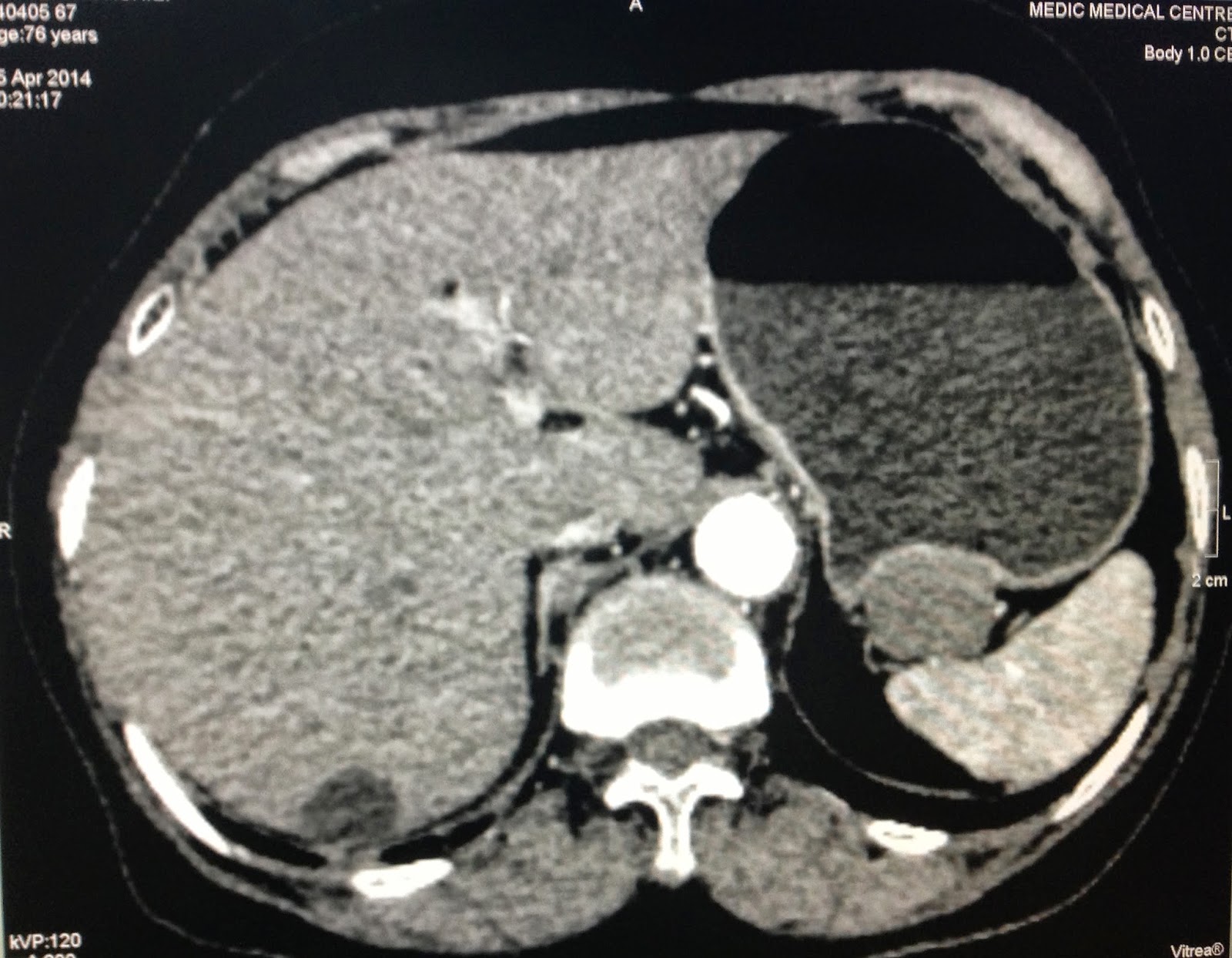
Woman 75 yo vomiting, endoscopy detected tumor extragastric fundus (see pictures).
Ultrasound of abdomen showed one hypoechoic mass with size of 4 cm, well-bordered at the hilus of spleen ( see 2 ultrasound pictures).
MSCT with CE found out this mass bending the wall of great curvature of stomach, very slow CE enhancement (see 3 CT pictures).
Blood tests of all markers are normal.
What is your suggestion of diagnosis?


