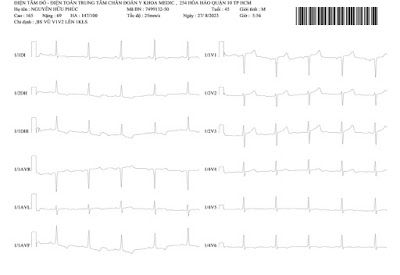
A 47 yo HTA male patient with feelings of heavy chest. His history notes 2 times of syncope and suffering from HTA since his ages of seventeen.
Chest X-RAY film is normal and so his EKG is ischemic heart disease.

Lab data is normal.
At last, DSA detects 2 renal arteries each side that belongs to a renal artery malformation.
REFERENCE
Renal arteries are a pair of lateral branches from abdominal aorta. Normally each kidney receives one renal artery. However, accessory renal arteries can also exist. The normal renal arteries enter the kidney through its hilum where as the accessory renal arteries might enter the renal artery through the hilum or through the surfaces of the kidney. Knowledge of the variations in the renal arteries is important for urologists, radiologists and surgeons in general.
Accessory renal arteries are common in 20–30% of individuals, usually arising from the aorta above or below the main renal artery. The variation in the number of arteries is because of persistence of lateral splanchnic arteries or due to the persistence of blood supply from lower level than normal.


