A 21 year-old female patient herself detects a small mass of right breast from years, but it is getting bigger recently for some months, hard feeling when palpation and painless. The skin of right breast is still normal and no axillary lymph node.
On ultrasound, the right breast tumor # 60x70 mm is in central, well capsulated, lobulated margins, ovoid, hypoechoic with many echo poor bands / clefts from central to peripheral tumor, medium vascularized.
MRI detects medium signal on T1W1, high on T2 STIR, contrast well captured, categoried type 2.
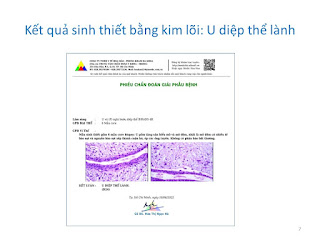
Result of core biopsy is a benign phyllodes tumor of the breast (PTB).
On the surface the tumor is nodular, while on section tumor is lobulated, solid in gray and gray-yellow color.
PTB is a very rare breast tumor in women aged 35 to 55 years. Our patient is younger but the progress of the tumor is the same in the literature: "unilateral, nodular, painless mass which has a history of the mass but that grows rapidly in the short term".


