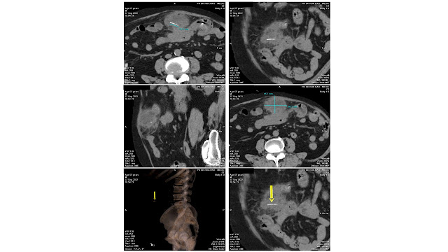
A 67 year-old male patient, presented with periumbilical and left lumbar pain for one month that was not response to treatment.
Abdominal ultrasound detected one mixed echogenic mass in the left lumbar mesentery with the diameter of 84 x 47mm. The conclusion was: Suspected mesenteric infarction (Differential diagnosis: Intra-abdominal Abscess) – Hepatic Steatosis – Abdominal Aortic Atherosclerosis.
MSCT of the abdomen showed a foreign object similar to a toothpick near the abdominal wall, right above the umbilicus, with a lenghth of 21 mm. The greater omentum surrounded the foreign object forming a mass with the diameters of 60 x 45 mm.
During operation, surgeons removed a foreign object which was highly suspected as a fish bone after dissecting the abscess in the greater omentum. The two adhering loops of small intestines were separated and reinforced with stitches.
Conclusion: Physicians should be on high alert when patients with abdominal pain not responding to the treatment. Abdominal ultrasound and MSCT help guiding the appropriate diagnosis for the case.


