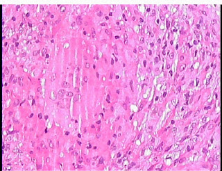
Female patient 32 yo, loss of weight #10kg with epigastric pain and nausea for 2 months.
She herself took gastric drugs for a while but failed so went to Medic for reexamination.
Ultrasound of abdomen at Medic revealed many lymph nodes that were suspected metastastic nodes and mesenteric thickening. Stomach walls were infiltratedly thickening with slighly splenomegaly.
Gastric endoscopy showed gastric body part inflammed roughly. Results of biopsy were TB inflammed of submucosa layer of stomach and chronic inflammation of duodenum.
MSCT confirmed that existed a lot of lymph nodes at hepatic hilus, lesser curvature of stomach, around celiac axis. These nodes maybe belong to TB nodes.
Result of biopsy of intraabdominal lymph node was TB inflammed nodes.
Discussions and Conclusions
TB of stomach is still a rare entity, which is about 1-2% of GI tract tuberculosis and in 0.5% of TB patients. Usually it is in secondary phase of the pulmonary TB disease whenever might happen in the past.
Our stomach TB [antrum and pyloric parts] patient is now getting better status, gained more 2 kg of weight while was taken TB drugs for 2 months of 6 month planned therapy.


