.
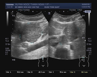
Female child 11 yo, with abdominal pain and diarrhoe 1/2 day.
Abdominal ultrasound detected a solid tumor # 45X48X52mm between liver and stomach which is look like hepatic tissue.
MRI with contrast shows tumor from left lobe of liver #50x42mm, regular boder, with hepatic signals, strong enhancement in arterial phase and wash out same liver tissue in late phase. A FNH in left liver lobe was been made in diagnostic.
Blood tests=
Open surgery to remove tumor for the child .
HISTOPATHLOGY RESULT=
REFERENCE=


