Man 55 yo for check- up MSCT total body and blood tests.
Radiologist report detected one mass # 1.6 cm at right lobe of thyroid gland, with HU 91 UI in comparison to the thyroid tissue background =121UI ( CT1 CT2 CT3).
Blood test is normal thyroid function.
Ultrasound scanning is second look:
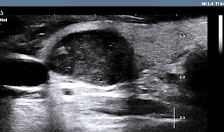
US 1: crossed-section of the hypoechoic focal lesion, well-bordered # 1.5 cm.
US 2: longitudinal scanning of the mass is 1. 7 cm, hypoechoic pattern, with
US 3: CDI = hypovascular mass, no lymph node in the neck.
This mass is in TI-RADS 3, need FNAC.
FNAC report suspected papillary carcinoma ( PTC).
Operation is done for subtotal thyroidectomy (see macro 1,2).


